. 1998 May;180(9):2442-9.
doi: 10.1128/JB.180.9.2442-2449.1998.

Isolation of an Escherichia coli K-12 mutant strain able to form biofilms on inert surfaces: involvement of a new ompR allele that increases curli expression

Affiliations

Isolation of an Escherichia coli K-12 mutant strain able to form biofilms on inert surfaces: involvement of a new ompR allele that increases curli expression

O Vidal et al. J Bacteriol. 1998 May.

Abstract

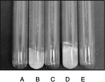
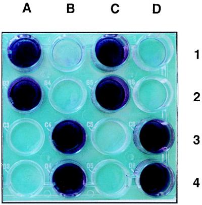
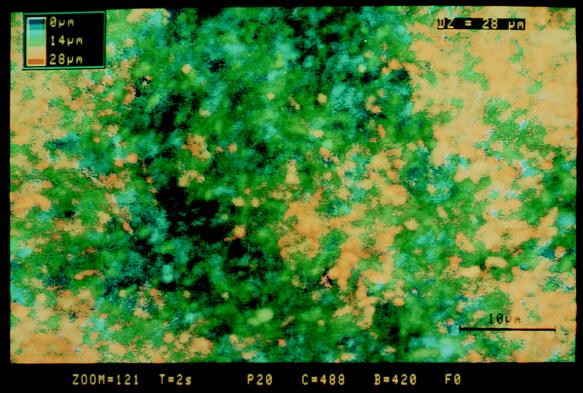
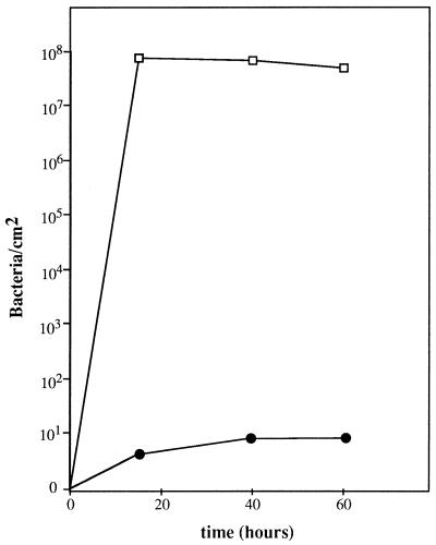
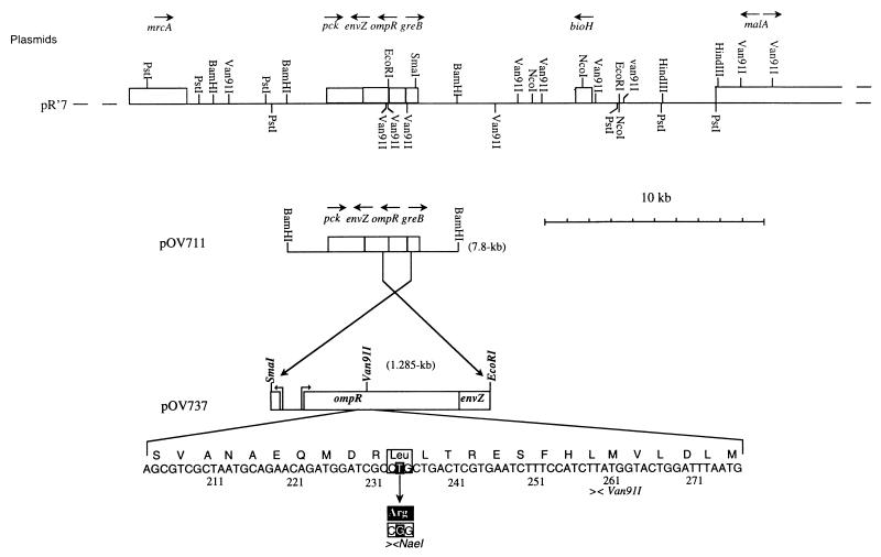
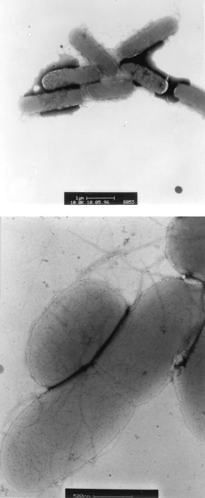
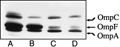
Classical laboratory strains of Escherichia coli do not spontaneously colonize inert surfaces. However, when maintained in continuous culture for evolution studies or industrial processes, these strains usually generate adherent mutants which form a thick biofilm, visible with the naked eye, on the wall of the culture apparatus. Such a mutant was isolated to identify the genes and morphological structures involved in biofilm formation in the very well characterized E. coli K-12 context. This mutant acquired the ability to colonize hydrophilic (glass) and hydrophobic (polystyrene) surfaces and to form aggregation clumps. A single point mutation, resulting in the replacement of a leucine by an arginine residue at position 43 in the regulatory protein OmpR, was responsible for this phenotype. Observations by electron microscopy revealed the presence at the surfaces of the mutant bacteria of fibrillar structures looking like the particular fimbriae described by the Olsén group and designated curli (A. Olsén, A. Jonsson, and S. Normark, Nature 338:652-655, 1989). The production of curli (visualized by Congo red binding) and the expression of the csgA gene encoding curlin synthesis (monitored by coupling a reporter gene to its promoter) were significantly increased in the presence of the ompR allele described in this work. Transduction of knockout mutations in either csgA or ompR caused the loss of the adherence properties of several biofilm-forming E. coli strains, including all those which were isolated in this work from the wall of a continuous culture apparatus and two clinical strains isolated from patients with catheter-related infections. These results indicate that curli are morphological structures of major importance for inert surface colonization and biofilm formation and demonstrate that their synthesis is under the control of the EnvZ-OmpR two-component regulatory system.


Figures

FIG. 1
FIG. 1
Biofilm development on the walls of glass test tubes containing β63 (A), RL101 (B), MG1655 (C), PHL628 (D), and no bacteria (control) (E). After inoculation, the tubes were gently shaken at 30°C. After 24 h, the medium (M63) was removed and the tubes were dried for 1 h at room temperature.
FIG. 2
FIG. 2
Biofilm development in the wells of a polystyrene microtiter plate. The wells contained RL101 (A1), β63 (B1), PHL628 (MG1655 adr-101) (C1), MG1655 (D1), PHL644 (MC4100 adr-101) (A2), PHL645 (MC4100 adr+) (B2), MG1655(pR′7) (C2), MG1655(pULB110) (D2), TK821 (MC4100 ompR331::Tn10) (A3), MC4100(pR′7) (B3), MC4100(pAT003) (C3), MC4100(pOV711) (D3), PHL745 (MC4100 adr-101 csgA::kan) (A4), PHL745(pCSG4) (B4), MC4100 csgA::kan(pCSG4) (C4), and MC4100(pOV737) (D4). The wells were filled with M63 medium. After inoculation, the plate was incubated for 48 h at 30°C. The liquid was removed from each well, and a drop of crystal violet was added to intensify the contrast. The wells were then washed twice (with 10 mM MgSO4), and the plate was dried.
FIG. 3
FIG. 3
3-D reconstitution of the biofilm developed by the adherent strain PHL628 on a glass coverslip. A test tube containing 3 ml of M63 medium was inoculated with the bacteria, and a glass coverslip was introduced into the tube. After being cultured for 24 h at 30°C, the slide was removed, stained with acridine orange, and observed by confocal laser microscopy (see Materials and Methods). Horizontal optical sections were collected at 1.0-μm intervals, digitized, and serially arranged to create 3-D reconstitutions. Pseudocolors were attributed to each point according to its distance from the glass surface. The color-to-distance conversion scale is shown at the upper left corner of the micrograph. Bar, 10 μm.
FIG. 4
FIG. 4
Quantification of biofilm development on the surface of polystyrene petri plates. Colonization by the adr-101 strain PHL644 (open squares) and the wild-type strain PHL645 (solid circles) is shown. The plates contained liquid M63 medium and were incubated at 30°C. The number of bacteria in the biofilm was measured as described in Materials and Methods.
FIG. 5
FIG. 5
Restriction maps of the chromosomal fragments cloned in this work and sequence of the ompR234 mutation. The ompR region of PHL628 was cloned in vivo on pR′7 (see text). A 7.8-kb BamHI fragment and a 1.285-kb SmaI-EcoRI fragment conferring the adherent phenotype were successively subcloned into pBR325 (to generate plasmids pOV711 and pOV737). Part of the nucleotide sequence of ompR (32) is also shown. The single-base pair mutation is indicated by a black square, with the corresponding modification of the amino acid sequence given in the solid box. The mutation created a NaeI restriction site (see text).
FIG. 6
FIG. 6
Electron micrographs of negatively stained bacteria of the biofilm-forming mutant PHL628. Bars, 1 μm (top) and 0.5 μm (bottom). The cells were cultivated in M63 medium at 30°C for 24 h.
FIG. 7
FIG. 7
Outer membrane proteins of the ompR234 strain PHL644 (lanes A and C) and the wild-type strain PHL645 (lanes B and D). Bacteria were grown to mid-log phase at 30°C in MOPS medium (lanes A and B) or in MOPS medium with 300 mM NaCl (lanes C and D). Equal amounts of each membrane fraction protein were separated by 6 M urea–SDS-PAGE.

Similar articles

Cited by

References

    1. Baker T A, Howe M M, Gross C A. Mu dX, a derivative of Mu d1 (lac Apr) which makes stable lacZ fusions at high temperature. J Bacteriol. 1983;156:970–974. - PMC - PubMed
    1. Bardonnet N, Blanco C. uidA antibiotic resistance cassettes for insertion mutagenesis, gene fusions and genetic constructions. FEMS Microbiol Lett. 1992;93:243–248. - PubMed
    1. Berlyn M K B, Low K B, Rudd K E. Linkage map of Escherichia coli K-12, edition 9. In: Neidhardt F C, Curtiss III R, Ingraham J L, Lin E C C, Low K B, Magasanik B, Reznikoff W S, Riley M, Schaechter M, Umbarger H E, editors. Escherichia coli and Salmonella: cellular and molecular biology. 2nd ed. Washington, D.C: American Society for Microbiology; 1996. pp. 1715–1902.
    1. Caldwell D E, Korber D R, Lawrence J R. Confocal laser microscopy and digital image analysis in microbial ecology. Adv Microb Ecol. 1992;12:1–67.
    1. Costerton J W, Lewandowski Z, De Beer D, Caldwell D, Korber D, James G. Biofilms, the customized microniche. J Bacteriol. 1994;176:2137–2142. - PMC - PubMed

Publication types

MeSH terms

LinkOut - more resources