. 1998 Jun;72(6):4560-70.
doi: 10.1128/JVI.72.6.4560-4570.1998.

The Us9 gene product of pseudorabies virus, an alphaherpesvirus, is a phosphorylated, tail-anchored type II membrane protein

Affiliations

The Us9 gene product of pseudorabies virus, an alphaherpesvirus, is a phosphorylated, tail-anchored type II membrane protein

A D Brideau et al. J Virol. 1998 Jun.

Abstract

The Us9 gene is highly conserved among the alphaherpesviruses sequenced to date, yet its function remains unknown. In this report, we demonstrate that the pseudorabies virus (PRV) Us9 protein is present in infected cell lysates as several phosphorylated polypeptides ranging from 17 to 20 kDa. Synthesis is first detected at 6 h postinfection and is sensitive to the DNA synthesis inhibitor phosphonoacetic acid. Unlike the herpes simplex virus type 1 Us9 homolog, which was reported to be associated with nucleocapsids in the nuclei of infected cells (M. C. Frame, D. J. McGeoch, F. J. Rixon, A. C. Orr, and H. S. Marsden, Virology 150:321-332, 1986), PRV Us9 localizes to the secretory pathway (predominately to the Golgi apparatus) and not to the nucleus. By fusing the enhanced green fluorescent protein (EGFP) reporter molecule to the carboxy terminus of Us9, we demonstrated that Us9 not only is capable of targeting a Us9-EGFP fusion protein to the Golgi compartment but also is able to direct efficient incorporation of such chimeric molecules into infectious viral particles. Moreover, through protease digestion experiments with Us9-EGFP-containing viral particles, we demonstrated that the Us9 protein is inserted into the viral envelope as a type II, tail-anchored membrane protein.


Figures

FIG. 1
FIG. 1
Us9 amino acid sequence and gene constructs. (A) Amino acid sequence of the Us9 open reading frame. The two in-frame methionine residues (#), the potential tyrosine kinase phosphorylation sites (∗), and the potential casein kinase I and II sites (∧) are indicated. The potential N-linked glycosylation (NXS/T) sequence is double underlined. The putative transmembrane domain is underlined, and the surrounding basic residues are indicated (+). (B) Diagrams of all the Us9 constructs used in this study. The two in-frame methionine residues (M), the putative transmembrane domain (TM), and the clusters of basic charges (+) are marked. The Us9 protein fusions with GST, the influenza A virus HA epitope, and EGFP are indicated. All constructs depicted are under the transcriptional control of the CMV immediate-early promoter.
FIG. 2
FIG. 2
Us9 protein in PRV Be- and Ba-infected cell lysates and virions. Monolayers of PK15 cells were infected with PRV Be or Ba virus at an MOI of 10. PRV-infected monolayers were radiolabeled for 11 h beginning at 5 h postinfection. (A) Cell lysates were either denatured with SDS and dithiothreitol or left untreated prior to immunoprecipitation with Us9 antiserum or preimmune serum. The identity of the polypeptide in nondenatured Be lysates immunoprecipitated with either Us9 antiserum or preimmune serum (indicated by an asterisk) is currently unknown. (B) Radiolabeled PRV virions were collected at 16 h postinfection by centrifugation through a 30% sucrose cushion and subjected to immunoprecipitation with Us9 antiserum. Positions of molecular mass markers (kilodaltons) are indicated on the left.
FIG. 3
FIG. 3
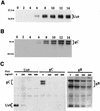
Analysis of Us9 protein kinetics in PRV Be-infected cell lysates. (A and B) Monolayers of PK15 cells were infected with PRV Be (MOI = 10), and cellular extracts were prepared at 0, 2, 4, 6, 8, 10, 12, and 14 h after infection. Ten micrograms of total cell lysate per lane was fractionated on an SDS–12.5% polyacrylamide gel, transferred to nitrocellulose, and Western blotted with either Us9 (A) or gC (B) antiserum. (C) For PAA treatment, 0, 200, 400, or 800 μg of PAA per ml was added to PK15 cells 1 h prior to and during viral infection. The cells were harvested and lysed at 10 h postinfection, and the cell lysate (10 μg) was analyzed by Western blotting with Us9, gC, or gB antiserum. The migration of molecular mass markers is indicated on the left in kilodaltons.
FIG. 4
FIG. 4
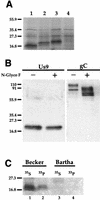
Us9 posttranslational modifications. (A) In vitro translation of Us9, Us9 M9A, and Us9-HA-tagged constructs. Plasmids pAB7 (lane 1), pAB19 (lane 2), and pAB11 (lane 3) were transcribed and translated in vitro in the presence of [35S]methionine by a rabbit reticulocyte lysate procedure. The bands migrating at approximately 27 and 35 kDa represent nonspecific translation products, as they are also detected during in vitro translation of the parental vector alone (pcDNA1/Amp) (lane 4). (B) N-glycosidase F treatment of Us9. PRV Be-infected cell lysates were incubated in either the presence (+) or absence (−) of N-glycosidase F (N-Glyco F). The treated lysates were separated on an SDS–12.5% polyacrylamide gel, electroblotted onto nitrocellulose, and analyzed by Western blotting with Us9 or gC antiserum. (C) Analysis of Us9 phosphoforms. PRV Be (lanes 1 and 2)- or Ba (lanes 3 and 4)-infected PK15 cells were radiolabeled overnight in the presence of either [35S]methionine-cysteine (lanes 1 and 3) or [33P]orthophosphate (lanes 2 and 4). Cellular extracts were prepared at 16 h postinfection and subjected to immunoprecipitation with Us9 antiserum. All of the immunoprecipitated products were analyzed by electrophoresis on an SDS–12.5% polyacrylamide gel followed by autoradiography. Positions of molecular mass markers (kilodaltons) are indicated on the left.
FIG. 5
FIG. 5
Immunofluorescence analysis of Us9 in PRV Be-infected cells. PK15 cells grown on glass coverslips to approximately 70% confluency were infected with PRV Be at an MOI of 10. At 2.5, 3, 4, and 6 h postinfection (hpi), the cells were fixed with formaldehyde and processed to detect Us9 protein by indirect immunofluorescence and confocal microscopy. A fluorescein isothiocyanate-conjugated donkey anti-rabbit immunoglobin G secondary antibody was used to visualize the Us9 antigen-antibody complexes.
FIG. 6
FIG. 6
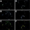
Colocalization of Us9 with envelope proteins gB and gC. PK15 cells grown on glass coverslips were infected with PRV Be at an MOI of 10. The cells were fixed and stained for Us9 and gB (A, B, and C) at 6 h postinfection and for Us9 and gC (D, E, and F) at 4 h post-infection. Us9 (A and D) was detected with an indocarbocyanine-conjugated (Cy3) donkey anti-rabbit secondary antibody. gB (B) and gC (E) were visualized with a fluorescein isothiocyanate-conjugated donkey anti-goat secondary antibody. The Us9 (red) and gB (green) colocalization is shown in panel C. The Us9 (red) and gC (green) colocalization is shown in panel F.
FIG. 7
FIG. 7
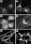
Transfection of Us9 constructs. PK15 cells were grown on glass coverslips and transfected by the calcium phosphate method with pAB7 (A), pBB14 (B, D, E, and F), or EGFP (C). At 72 h posttransfection, Us9 was detected by indirect immunofluorescence microscopy with Us9 antiserum (A) as described in the legend to Fig. 5. The localization of pBB14 (B, D, E, and F) and EGFP (C) was determined at 48 h posttransfection by confocal microscopy under UV illumination. Panel D is a higher magnification of the Us9-EGFP-transfected cells shown in panel B, clearly demonstrating expression of the fusion protein in the Golgi compartment and plasma membrane. (E and F) Sensitivity of Us9-EGFP (pBB14) to BFA treatment. PK15 cells transfected with pBB14 were treated with 2.5 μg of BFA per ml for 60 minutes (E). Some BFA-treated cells were then washed three times with fresh medium and allowed to recover for 1 h (F).
FIG. 8
FIG. 8
Us9 is a type II membrane protein. Us9-EGFP-containing virions were isolated from the medium of Us9-EGFP-expressing cells infected with PRV Be or Ba (MOI = 10) as described in Materials and Methods. The purified PRV Be virions (A) or Ba virions (B and C) were treated with (lanes 3 and 4) or without (lanes 1 and 2) proteinase K in either the presence (lanes 2 and 4) or absence (lanes 1 and 3) of 1% NP-40. The samples were then fractionated on an SDS–12.5% polyacrylamide gel and analyzed by Western blotting with either Us9 (A and B) or GFP (C) antiserum. Positions of molecular mass markers (kilodaltons) are indicated on the left.

Similar articles

Cited by

References

    1. Babic N, Klupp B, Brack A, Mettenleiter T C, Ugolini G, Flamand A. Deletion of glycoprotein gE reduces the propagation of pseudorabies virus in the nervous system of mice after intranasal inoculation. Virology. 1996;219:279–284. - PubMed
    1. Brandimarti R, Roizman B. Us9, a stable lysine-less herpes simplex virus 1 protein, is ubiquitinated before packaging into virions and associates with proteosomes. Proc Natl Acad Sci USA. 1997;94:13973–13978. - PMC - PubMed
    1. Browne H, Bell S, Minson T, Wilson D W. An endoplasmic reticulum-retained herpes simplex virus glycoprotein H is absent from secreted virions: evidence for reenvelopment during egress. J Virol. 1996;70:4311–4316. - PMC - PubMed
    1. Card J P, Rinaman L, Schwaber J S, Miselis R R, Whealy M E, Robbins A K, Enquist L W. Neurotropic properties of pseudorabies virus: uptake and transneuronal passage in the rat central nervous system. J Neurosci. 1990;10:1974–1994. - PMC - PubMed
    1. Card J P, Whealy M E, Robbins A K, Enquist L W. Pseudorabies virus envelope glycoprotein gI influences both neurotropsim and virulence during infection of the rat visual system. J Virol. 1992;66:3032–3041. - PMC - PubMed

Publication types

LinkOut - more resources