. 1998 Jun;72(6):4580-8.
doi: 10.1128/JVI.72.6.4580-4588.1998.

A chicken embryo eye model for the analysis of alphaherpesvirus neuronal spread and virulence

Affiliations

A chicken embryo eye model for the analysis of alphaherpesvirus neuronal spread and virulence

B W Banfield et al. J Virol. 1998 Jun.

Abstract

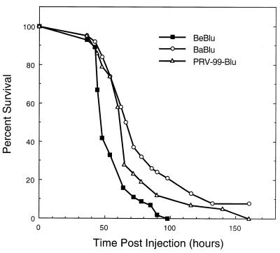
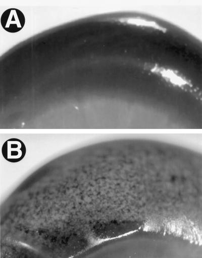
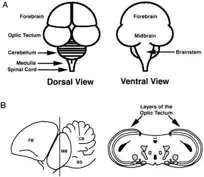
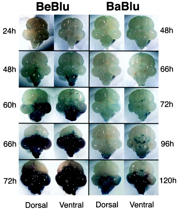
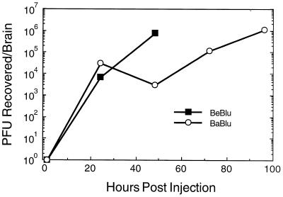
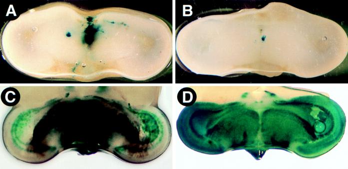
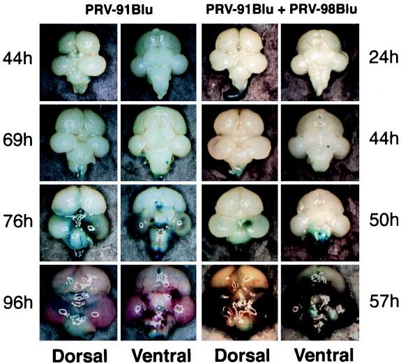
We describe use of developing chicken embryos as a model to study neuronal spread and virulence of pseudorabies virus (PRV). At embryonic day 12, beta-galactosidase-expressing PRV strains were injected into the vitreous humor of one eye, and virus replication and spread from the eye to the brain were measured by beta-galactosidase activity and the recovery of infectious virus from tissues. The wild-type PRV strain, Becker, replicated in the eye and then spread to the brain, causing extensive pathology characterized by edema, hemorrhage, and necrosis that localized to virally infected tissue. The attenuated vaccine strain, Bartha, replicated in the eye and spread throughout specific regions of the brain, producing little to no overt pathology. Becker mutants lacking membrane proteins gE or gI replicated in the eye and were able to spread to the brain efficiently. The pathology associated with replication of these mutants in the brain was intermediate to that induced by Becker or Bartha. Mixed infection of a gE deletion mutant and a gI deletion mutant restored the pathogenic phenotype to wild-type levels. These data indicate that the replication of virus in embryonic brain tissue is not sufficient to induce the characteristic pathological response and that the gE and gI gene products actively affect pathological responses in the developing chicken brain.


Figures

FIG. 1
FIG. 1
Survival curves of PRV strains in the chicken embryo eye model. Embryos were injected on E12 with 105 PFU of virus in a volume of 1 μl. Virus was delivered into the vitreous body of the right eye, using a 10-μl Hamilton syringe. Animal survival was monitored at least every 6 h over a 168-h period. The numbers of embryos used were 45, 38, and 43 for BeBlu, BaBlu, and PRV-99Blu, respectively.
FIG. 2
FIG. 2
Closeup images of the eyes from a PRV-infected embryo. The uninjected (A) and injected (B) eyes from a BaBlu-infected embryo were harvested at 72 h after infection.
FIG. 3
FIG. 3
Replication of PRV strains in the eye. At the indicated times, the inoculated eyes from three embryos were harvested and pooled, and infectious virus was released from tissue as described in Materials and Methods. Virus titer was determined by plaque assay on PK15 cells in duplicate.
FIG. 4
FIG. 4
Schematic drawing of the chicken embryo brain. (A) Dorsal and ventral views of the embryonic brain. (B) Left, lateral view of the chick brain. FB, forebrain; MB, midbrain; BS, brainstem; CB, cerebellum. Right, coronal section through the midbrain, the position of which is indicated on the left side by the dark line through the midbrain. Areas shaded in gray represent known retinorecipient regions in addition to the optic tectum. Areas shaded in black represent ventricles.
FIG. 5
FIG. 5
Progression of PRV infection in the chicken embryo brain. At the indicated times after injection into the right eye, the brains from infected embryos were removed and placed in X-Gal. Only infected tissue close to the surface of the brain or exposed by necrosis was stained blue. Dorsal and ventral views of each brain are shown. Note that the times that the brains were collected are different for Be-Blu- and Ba-Blu-infected embryos. It is important to note that many of the BeBlu-infected animals sacrificed at 48 h had more extensive infection than the examples illustrated and that in some cases their brains resembled the 66-h brain. This inherent variability is likely due to the use of genetically outbred animals but nevertheless emphasizes the speed with which BeBlu infection exerts its pathology.
FIG. 6
FIG. 6
Replication of PRV strains in the brain. At the indicated times after injection, brains from three infected embryos were removed and pooled, and infectious virus was released from tissue as described in Materials and Methods. Virus titer was determined by plaque assay on PK15 cells in duplicate.
FIG. 7
FIG. 7
PRV infection of the midbrain after intraocular inoculation. The brains of infected embryos were sliced in the coronal plane through the midbrain by using a razor blade and placed in X-Gal substrate buffer for 1 h. (A) Section through the midbrain of a BeBlu-infected embryo at 48 h postinfection; (B) section through the midbrain of a BaBlu-infected embryo at 48 h postinfection; (C) section through the midbrain of a BeBlu-infected embryo at 60 h postinfection; (D) section through the midbrain of a BaBlu-infected embryo at 96 h postinfection.
FIG. 8
FIG. 8
Progression of infection of gE and gI mutant PRV strains in the chicken embryo brain. At the indicated times after injection into the right eye, the brains from infected embryos were removed and placed in X-Gal. Only infected tissue close to the surface of the brain or exposed by necrosis was stained blue. Dorsal and ventral views of each brain are shown. PRV-91Blu + PRV-98Blu, coinfection experiment where equal amounts of the gE mutant PRV-91Blu and the gI mutant PRV-98Blu were mixed and injected. Note that the times that the brains were collected are different for the two infections.

References

    1. Anderson K. Pathogenesis of herpes simplex virus infection in chick embryos. Am J Pathol. 1940;16:137–156. - PMC - PubMed
    1. Babic N, Mettenleiter T C, Flamand A, Ugolini G. Role of essential glycoproteins gII and gp50 in transneuronal transfer of pseudorabies virus from the hypoglossal nerves of mice. J Virol. 1993;67:4421–4426. - PMC - PubMed
    1. Banfield, B. W., and L. W. Enquist. Unpublished observations.
    1. Banfield, B. W., C. Lee, S. Marks, N. Brecha, and L. W. Enquist. Unpublished observations.
    1. Bang F. Experimental infection of the chick embryo with the virus of pseudorabies. J Exp Med. 1942;76:263–269. - PMC - PubMed

Publication types

Substances

LinkOut - more resources