. 1998 Jun;72(6):4623-32.
doi: 10.1128/JVI.72.6.4623-4632.1998.

Loss of CD4+ T cells in human immunodeficiency virus type 1-infected chimpanzees is associated with increased lymphocyte apoptosis

Affiliations

Loss of CD4+ T cells in human immunodeficiency virus type 1-infected chimpanzees is associated with increased lymphocyte apoptosis

I C Davis et al. J Virol. 1998 Jun.

Abstract

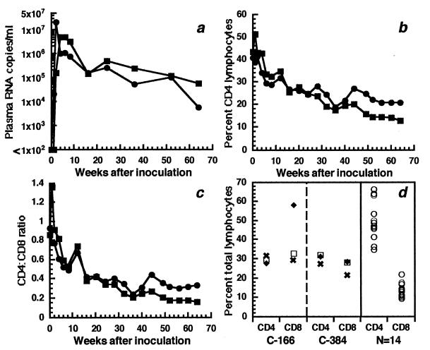
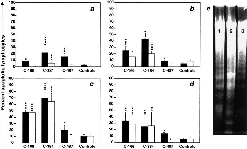
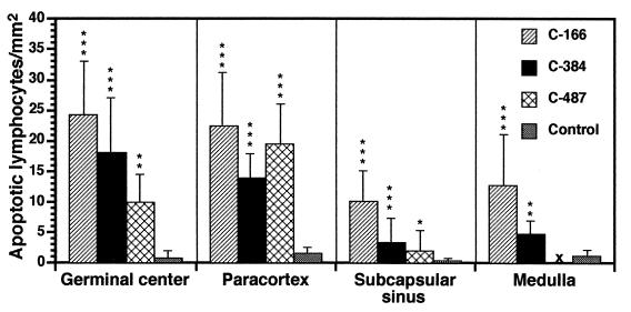
Supportive evidence that apoptosis contributes to loss of CD4+ lymphocytes in human immunodeficiency virus type 1 (HIV-1)-infected humans comes from an apparent lack of abnormal apoptosis in apathogenic lentivirus infections of nonhuman primates, including HIV-1 infection of chimpanzees. Two female chimpanzees were inoculated, one cervically and the other intravenously, with HIV-1 derived from the LAI/LAV-1b strain, which was isolated from a chimpanzee infected with the virus for 8 years. Within 6 weeks of infection, both recipient chimpanzees developed a progressive loss of CD4+ T cells which correlated with persistently high viral burdens and increased levels of CD4+ T-cell apoptosis both in vitro and in vivo. Lymph nodes from both animals also revealed evidence of immune hyperactivation. Intermediate levels of T-cell apoptosis in both peripheral blood and lymph nodes were seen in a third chimpanzee that had been infected with the LAI/LAV-1b strain for 9 years; this animal has maintained depressed CD4/CD8 T-cell ratios for the last 3 years. Similar analyses of cells from 4 uninfected animals and 10 other HIV-1-infected chimpanzees without loss of CD4+ cells revealed no difference in levels of apoptosis in these two control groups. These results demonstrate a correlation between immune hyperactivation, T-cell apoptosis, and chronic loss of CD4+ T cells in HIV-1-infected chimpanzees, providing additional evidence that apoptosis is an important factor in T-cell loss in AIDS. Furthermore, the results show that some HIV-1 strains are pathogenic for chimpanzees and that this species is not inherently resistant to HIV-1-induced disease.


Figures

FIG. 1
FIG. 1
Evaluation of chimpanzees C-166 (▪) and C-384 (•) after cervical and intravenous inoculation, respectively, of HIV-1JC499. (a) Plasma HIV-1 RNA levels. (b) Percentages of CD4+ T cells in peripheral blood. (c) CD4/CD8 ratios in peripheral blood. (d) Percentages of CD4+ and CD8+ T cells in peripheral lymph nodes. For C-166 and C-384, the LN biopsies were done 12 (✖), 24 (□), and 49 (⧫) weeks after inoculation. All of the control LN biopsies were performed on chimpanzees infected with different HIV-1 strains for various times.
FIG. 2
FIG. 2
Three-color FACS analysis of CD4+ and CD8+ cells in PBMC samples from nonprogressor chimpanzee C-460 (a to c) and progressor C-166 (d to f) after culture for 48 h in the absence of mitogens. (a and d) Gates for 7-AADNEG live and 7-AADLO apoptotic cells in the FL-3 channel. (b and e) Percentages of CD4+ and CD8+ T cells in the 7-AADNEG population. (c and f) Percentages of apoptotic CD4+ and CD8+ T cells in the 7-AADLO population. In this example, the percentage of apoptotic CD4+ T cells in C-460’s PBMC is 5% (2 ÷ 39), and that in C-166’s PBMC is 22% (5 ÷ 23).
FIG. 3
FIG. 3
Percentages of apoptotic CD4+ (closed bars) and CD8+ (open bars) T cells in PBMC samples from progressor and control (nonprogressor and uninfected) chimpanzees (n = 8). (a) Ex vivo apoptosis. (b) In vitro apoptosis after culture for 48 h in medium alone. (c and d) In vitro apoptosis after culture for 48 h in the presence of PHA (10 μg/ml) or SEB (1 μg/ml), respectively. Values for the three progressor chimpanzees are means of four different PBMC samples collected at 1-month intervals. Error bars represent two standard errors of the mean. ∗, P < 0.05; ∗∗, P < 0.005; ∗∗∗, P < 0.0005 (all relative to control values). (e) Agarose gel electrophoresis of low-molecular-weight DNA in PBMC from C-166 (lane 1), C-384 (lane 2), and C-487 (lane 3) after culture for 48 h in medium alone.
FIG. 4
FIG. 4
Correlation between ex vivo apoptosis of CD4+ T cells and in vivo CD4+ T-cell loss in HIV-1-infected chimpanzees: ▪, C-166; •, C-384; ▴, C-487; ✖, nonprogressors (those analyzed by FACS; see Table 1). The slope of the line, the correlation coefficient (r2), and the P value were determined by linear regression; placement of the line is an approximation.
FIG. 5
FIG. 5
Immune hyperactivation and lymphocyte apoptosis in LN from progressor chimpanzees. (a) Follicular hyperplasia in a hematoxylin-eosin-stained LN section from C-384. Magnification, ×40. (b) Histology of normal LN tissue from nonprogressor C-534. Magnification, ×40. (c) TUNEL-positive lymphocytes in germinal center of LN from C-166. Magnification, ×100. (d) TUNEL-positive lymphocytes in paracortex of LN from C-384. Magnification, ×1,000. (e) Tingible body macrophage containing multiple phagocytosed apoptotic nuclei in germinal center of LN from C-166. Magnification, ×1,000. (f) TUNEL-stained LN tissue from nonprogressor C-304. Magnification, ×400. (g and h) Transmission electron micrograph of LN biopsy tissue from C-384, showing apoptotic lymphocytes with highly condensed chromatin, cytoplasmic condensation, and plasmalemmal zeiosis. Magnification, ×27,000.
FIG. 6
FIG. 6
TUNEL-positive (apoptotic) lymphocytes in major histoarchitectural regions of LN tissue from progressor and control (nonprogressor and uninfected) chimpanzees. Three consecutive tissue sections were analyzed for each LN biopsy. ×, not done due to lack of available tissue. ∗, P < 0.05; ∗∗, P < 0.005; ∗∗∗, P < 0.0005. Error bars represent two standard errors of the mean.

Similar articles

Cited by

References

    1. Ameisen J C. From cell activation to cell depletion. The programmed cell death hypothesis of AIDS pathogenesis. In: Andrieu J-M, Lu W, editors. Cell activation and apoptosis in HIV infection. New York, N.Y: Plenum Press; 1995. pp. 139–163. - PubMed
    1. Ameisen J C, Capron A. Cell dysfunction and depletion in AIDS: the programmed cell death hypothesis. Immunol Today. 1991;12:102–105. - PubMed
    1. Antoni B A, Sabbatini P, Rabson A B, White E. Inhibition of apoptosis in human immunodeficiency virus-infected cells enhances virus production and facilitates persistent infection. J Virol. 1995;69:2384–2392. - PMC - PubMed
    1. Ascher M S, Sheppard H W. AIDS as immune system activation: a model for pathogenesis. Clin Exp Immunol. 1988;73:165–167. - PMC - PubMed
    1. Badley A D, Dockrell D, Simpson M, Schut R, Lynch D H, Leibson P, Paya C V. Macrophage-dependent apoptosis of CD4+ T lymphocytes from HIV-infected individuals is mediated by FasL and tumor necrosis factor. J Exp Med. 1997;185:55–64. - PMC - PubMed

Publication types