. 1998 Jun;72(6):4686-93.
doi: 10.1128/JVI.72.6.4686-4693.1998.

Vpr stimulates viral expression and induces cell killing in human immunodeficiency virus type 1-infected dividing Jurkat T cells

Affiliations

Vpr stimulates viral expression and induces cell killing in human immunodeficiency virus type 1-infected dividing Jurkat T cells

X J Yao et al. J Virol. 1998 Jun.

Abstract

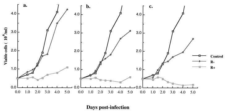
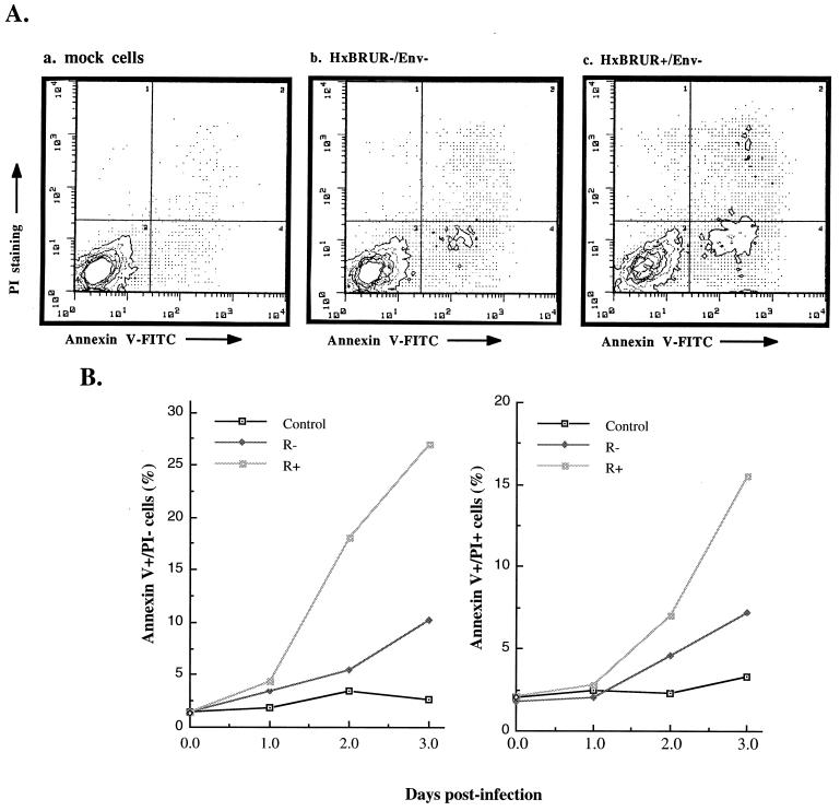
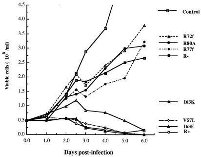
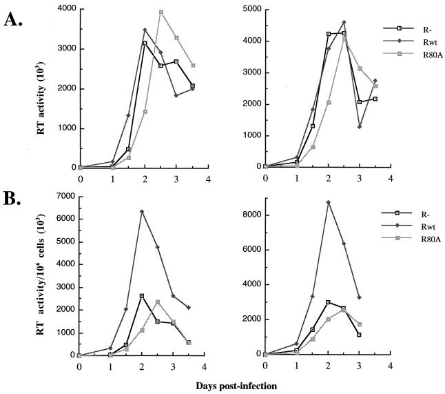
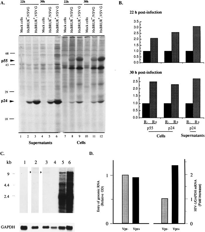
In this study we investigated the effects of Vpr during human immunodeficiency virus (HIV) infection of proliferating Jurkat T cells by using a vesicular stomatitis virus envelope G glycoprotein pseudotyped HIV superinfection system. We observe that the expression of Vpr results in a severe reduction in the life span of HIV type 1 (HIV-1)-infected dividing T cells in culture. In agreement with a recent report (S. A. Stewart, B. Poon, J. B. M. Jowett, and I. S. Chen, J. Virol. 71:5579-5592, 1997), we show that events characteristic of apoptotic cell death are involved in the Vpr-mediated cytopathic effects. Our results also show that infection with viruses expressing the wild-type vpr gene results in an increase in viral gene expression and production. Interestingly, the effects of Vpr on cell viability and on viral gene expression both correlate with the ability of the protein to induce a cell cycle arrest in the G2/M phase. Mutagenesis analyses show that the C terminus of Vpr is essential for these biological activities. Although the role of Vpr is currently associated with the infection of nondividing cells, our results suggest that Vpr can also directly increase viral replication in vivo in infected dividing T cells. Furthermore, these in vitro observations suggest that Vpr-mediated cytotoxic effects could contribute to the CD4+ depletion associated with AIDS progression.


Figures

FIG. 1
FIG. 1
Vpr increases cell killing during single-cycle infection of dividing Jurkat T cells. Jurkat T cells were mock infected or infected with VSV-G pseudotyped Vpr+ and Vpr HIV-1 viruses at MOIs of 5, 10, and 20 (respectively presented in panels a, b, and c). At different time intervals after infection, the number of viable cells was counted by trypan blue exclusion assay. The values represented means from duplicate samples. Similar data were obtained in two independent experiments.
FIG. 2
FIG. 2
Vpr induces apoptosis during single-cycle infection of dividing Jurkat T cells. (A) Jurkat T cells were infected with VSV-G pseudotyped Vpr+ or Vpr HIV-1 viruses. At 48 h p.i., the infected or mock-infected cells were costained with Annexin V and PI and analyzed by flow cytometry. The axes represent the cell-associated fluorescence intensity of Annexin V (x axis) and PI (y axis). (B) The percentage of Annexin-V+ PI+ or Annexin-V+ PI cells within the different infected cell populations were evaluated by Annexin V-PI costaining and flow cytometry analysis at 1, 2, and 3 days p.i.
FIG. 3
FIG. 3
Effects of different Vpr mutants on cell killing in Jurkat T cells. Induction of the cytopathic effect associated with different vpr alleles is shown. After infection with the indicated pseudotyped HIV-1 viruses, the number of viable cells in the cultures was determined at the indicated time intervals.
FIG. 4
FIG. 4
Vpr increases HIV viral production in dividing Jurkat T cells. Jurkat T cells were infected with VSV-G pseudotyped Vpr+, Vpr, or RR80A HIV-1 viruses at an MOI of 5 (left panels) or 10 (right panels). At each 12-h interval, the supernatants were collected and the virion-associated RT activities per volume of supernatants (RT activity/50 μl) (A) or the virion-associated RT activities relative to 106 viable cells (RT activity/50 μl/106 cells) (B) were determined. Values were representative of the data obtained from three independent experiments.
FIG. 5
FIG. 5
Vpr increases HIV viral gene expression in dividing Jurkat T cells. (A) Expression of viral protein in Jurkat T cells infected with VSV-G pseudotyped Vpr+ and Vpr HIV-1 viruses at MOIs of 10. At 22 and 30 h p.i., equivalent numbers of infected trypan blue-negative cells from different cultures were metabolically labeled with [35S]methionine. The viral proteins in both cell lysates and lysed supernatants were immunoprecipitated with an HIV-positive human serum and analyzed by SDS-polyacrylamide gel electrophoresis and autoradiography. (B) Densitometric analysis of the autoradiograms presented in panel A. The densitometric values of Gag p55 and p24 proteins from Vpr HIV-1-infected cells were arbitrarily set to 1. The densitometric values were highly reproducible. The standard deviation was less than 5%. (C) Expression of HIV-1 RNA in dividing Jurkat T cells. Jurkat cells were mock infected (lanes 1 and 4) or infected with Vpr (lanes 2 and 5) or Vpr+ (lanes 3 and 6) VSV-G pseudotyped HIV-1 particles at an MOI of 10 for 6 h. Cytoplasmic RNA was isolated from cells (lanes 1 to 3), or cells were cultured for an additional 18 h for steady-state mRNA measurements (lanes 4 to 6). RNA was fractionated, transferred to a nylon membrane, and probed sequentially with a 32P-labeled 285-bp probe for the HIV-1 5′ leader sequence and a 400-bp probe for GAPDH. The signals for internalized genomic RNA following the adsorption period are indicated by the arrowheads. (D) Results of densitometric scanning of internalized genomic RNA signals of panel C, lanes 2 and 3, and of the total of the spliced and unspliced RNA signals in lanes 5 and 6 of panel C. All signals were related to GAPDH. Relative optical density and fold increase in HIV-1/GAPDH mRNA were both related to the Vpr lane (set to 1). The densitometric values were highly reproducible. The standard deviation was less than 5%.

References

    1. Agostini I, Navarro J M, Rey F, Bouhamdan M, Spire B, Vigne R, Sire J. The human immunodeficiency virus type 1 Vpr transactivator: cooperation with promoter-bound activator domains and binding to TFIIB. J Mol Biol. 1996;261:599–606. - PubMed
    1. Ayyavoo V, Mahboubi A, Mahalingam S, Ramalingam R, Kudchodkar S, Williams W V, Green D R, Weiner D B. HIV-1 Vpr suppresses immune activation and apoptosis through regulation of nuclear factor κB. Nat Med. 1997;3:1117–1123. - PubMed
    1. Balliet J W, Kolson D L, Eiger G, Kim F M, McGann K A, Srinivasan A, Collman R. Distinct effects in primary macrophages and lymphocytes of the human immunodeficiency virus type 1 accessory genes vpr, vpu, and nef: mutational analysis of a primary HIV-1 isolate. Virology. 1994;200:623–631. - PubMed
    1. Bartz S R, Rogel M E, Emerman M. Human immunodeficiency virus type 1 cell cycle control: Vpr is cytostatic and mediates G2 accumulation by a mechanism which differs from DNA damage checkpoint control. J Virol. 1996;70:2324–2331. - PMC - PubMed
    1. Chun T-W, Chadwick K, Margolick J, Siliciano R F. Differential susceptibility of naive and memory CD4+ T cells to the cytopathic effects of infection with human immunodeficiency virus type 1 strain LAI. J Virol. 1997;71:4436–4444. - PMC - PubMed

Publication types

Substances