. 1998 Jun;72(6):4765-74.
doi: 10.1128/JVI.72.6.4765-4774.1998.

Mutations in the leucine zipper-like heptad repeat sequence of human immunodeficiency virus type 1 gp41 dominantly interfere with wild-type virus infectivity

Affiliations

Mutations in the leucine zipper-like heptad repeat sequence of human immunodeficiency virus type 1 gp41 dominantly interfere with wild-type virus infectivity

S S Chen et al. J Virol. 1998 Jun.

Abstract

It has been previously shown that a proline substitution for any of the conserved leucine or isoleucine residues located in the leucine zipper-like heptad repeat sequence of human immunodeficiency virus type 1 (HIV-1) gp41 renders viruses noninfectious and envelope (Env) protein unable to mediate membrane fusion (S. S.-L. Chen, C.-N. Lee, W.-R. Lee, K. McIntosh, and T.-M. Lee, J. Virol. 67:3615-3619, 1993; S. S.-L. Chen, J. Virol. 68:2002-2010, 1994). To understand whether these variants could act as trans-dominant inhibitory mutants, the ability of these mutants to inhibit wild-type (wt) virus infectivity was examined. Comparable amounts of cell- and virion-associated gag gene products as well as virion-associated gp41 were found in transfection with wt or mutant HIV-1 provirus. Viruses obtained from coexpression of wt provirus with mutant 566 or 580 provirus inhibited more potently the production of infectious virus than did viruses generated from cotransfection of wt provirus with other mutant proviruses. Nevertheless, all viruses produced from mixed transfection showed decreased infectivity compared with that of the wt virus when a multinuclear-activation beta-galactosidase induction assay was performed. The ability of wt Env to induce cytopathic effects was inhibited by coexpression with mutant Env. Coexpression of mutants inhibited the ability of the wt protein to mediate virus-to-cell transmission, as demonstrated by an env trans-complementation assay with a defective HIV-1 proviral vector. These observations indicated that mutant Env, per se, interferes with wt Env function. Moreover, cotransfection of wt and mutant proviruses produced amounts of cell- and virion-associated gag gene products comparable to those produced by transfection of wt provirus. Similar amounts of gp41 were also found in virions generated from wt-mutant cotransfection as well as from wt transfection alone. These results indicated that the inhibitory effect conferred by mutants on the wt virus infectivity does not involve the late steps of Gag protein assembly and budding, but they suggest that the wt and mutant Env proteins form a dysfunctional hetero-oligomer which is impaired in an early step of the virus replication cycle. Our study demonstrates that mutations in the HIV-1 gp41 leucine zipper-like heptad repeat sequence dominantly inhibit infectious virus production.


Figures

FIG. 1
FIG. 1
Schematic representation of the HIV-1 gp41 ectodomain. Amino acid residues are numbered according to their positions in the HXB2 Env protein. The N-terminal leucine zipper-like heptad repeat sequence is shown as the hatched arrow directed to the right, and its amino acid sequence in single-letter code is indicated. The conserved leucine and isoleucine residues located in this motif, which are numbered and indicated by boldface, were each replaced by a proline residue and examined in this study. The C-terminal α-helical sequence is illustrated as the hatched arrow directed to the left. This domain forms a heterodimer with the N-terminal zipper motif α helix, and three molecules of heterodimers fold into a six-stranded helical bundle. Within the bundle the three N-terminal helices constitute a central, parallel, trimeric coiled-coil structure, whereas the three C-terminal helices pack antiparallelly into the hydrophobic groves on the surface of the N-terminal trimer. The N-glycosylation sites (Ψ) and intramolecular disulfide bond (C—C) are also shown.
FIG. 2
FIG. 2
Expression of viral proteins encoded by wt and zipper motif mutant HXB2 proviruses. 293 monolayers were transfected by a standard calcium phosphate coprecipitation method with 10 μg each of wt or mutant HXB2 proviruses as indicated. The numbers shown in parentheses indicate the locations where the conserved leucine or isoleucine residues in the gp41 zipper motif were replaced by a proline residue. Mock-transfected cells were included as a negative control (lane 1). Two days after transfection, lysates of cells and virions were prepared as described in Materials and Methods. Equal aliquots of cell lysates and virus fractions were separated by SDS–10% PAGE, and proteins were visualized by Western immunoblotting analysis with rabbit anti-p25/p24 (strain SF2) (A), sheep anti-gp120 (B), or anti-gp41-specific Chessie 8 MAb (C).
FIG. 3
FIG. 3
Effect of cotransfection with wt and mutant HXB2 proviruses on production of infectious virus. 293 cells were transfected with 5 μg of wt HXB2 or 5 μg of wt HXB2 plus 10 μg of mutant HXB2 proviruses as indicated. The total amount of DNA in all transfections was kept constant at 15 μg by adding pHXBCATΔBgl plasmid DNA. Two days after transfection, culture supernatants were collected, centrifuged, and passed through 0.45-μm-pore-size membranes. Virions containing 5 × 10⁴ cpm of RT activity from each virus stock were used to challenge 2 × 10⁶ SupT1 cells. The cultures were monitored for virion-associated RT production at different times postinfection.
FIG. 4
FIG. 4
Env proteins encoded by wt and mutant pSVE7 plasmids. COS-1 cells were cotransfected by the DEAE-dextran method with 5 μg each of wt or mutant pSVE7 plasmid in the presence of 2 μg of pIIIextat. Two days after transfection, cell lysates were prepared and equal amounts of cell lysates were subjected to SDS-PAGE followed by Western blotting with sheep anti-gp120.
FIG. 5
FIG. 5
Inhibition of the wt Env-mediated cytopathic effects by coexpression with wt and mutant Env proteins. HeLa-CD4-LTR-β-gal cells grown in six-well plates were transfected with 1 μg of pIIIextat and 2 μg of wt pSVE7 in the presence or absence of 4 μg of mutant pSVE7 plasmids as indicated, using 10 μl of Superfect transfection reagent according to the Qiagen protocol. Transfection with pIIIextat was used as a negative control (A). DNA of pSVE7ΔKS was added to transfection mixtures to keep the total DNA amount in each transfection constant. Two days after transfection, cell cultures were photographed under a light microscope. Magnification, ×200.
FIG. 6
FIG. 6
Virus-to-cell transmission of a defective provirus mediated by wt and mutant Env coexpression. (A) wt/mutant pSVE7 DNA ratio of 1:1. 293 cells were cotransfected with 10 μg of pHXBCATΔBgl and 10 μg of wt pSVE7 or with 10 μg each of pHXBCATΔBgl, wt, and mutant pSVE7 plasmids as indicated. Transfection with the defective provirus alone was used as a control (lane 1). Plasmid SVE7ΔKS was added to transfection mixtures to make the total amount of DNA in each transfection constant. Two days after transfection, cell-free viruses were prepared, and virus from each stock containing 5 × 10⁴ cpm of RT activity was applied to subconfluent HeLa-CD4-LTR-β-gal cells grown in 60-mm-diameter petri dishes. Unbound viruses were removed after overnight incubation at 37°C, and fresh media were added to cultures. Three days after transfection, cell lysates were prepared and assayed for CAT activity. (B) wt/mutant pSVE7 DNA ratio of 1:2. Transfection was performed as described for panel A except that 7.5 μg of pHXBCATΔBgl, 5 μg of wt pSVE7, and 10 μg of various pSVE7 mutants were used in transfection. Viruses with 2 × 10⁵ cpm of RT activity were applied to HeLa-CD4-LTR-β-gal cells, and CAT activity was assayed 3 days after infection.
FIG. 7
FIG. 7
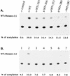
Effect of coexpression of wt and zipper motif mutant proteins on the wt Env-mediated virus-to-cell transmission. (A and B) Env protein expression. COS-1 cells were transfected with 5 μg each of pBaby, wt pBSX, or mutant pBSX, as indicated, by the DEAE-dextran method. Two days after transfection, equal amounts of cells lysates from each transfection were analyzed by Western blotting with anti-gp120 (A) or Chessie 8 anti-gp41 MAb (B). (C) Ability of mutant proteins to inhibit wt Env-induced virus entry into CD4+ cells. COS-1 cells were transfected with 5 μg of pHXBCATΔBgl and 2 μg of wt pBSX in the presence or absence of 2 μg of mutant pBSX as indicated. Transfection with pHXBCATΔBgl and pBaby was used as a negative control (lane 1). The total DNA amount in all transfections was kept constant by adding pBaby DNA. Two days following transfection, cell-free viruses were prepared, and 105 cpm of RT activity from each virus stock was then applied to HeLa-CD4-LTR-β-gal cells. Three days after infection, cell lysates were prepared and assayed for CAT activity.
FIG. 8
FIG. 8
Interference conferred by zipper motif Env mutants with virus-to-cell transmission assayed in PM1 cells. Recombinant viruses were generated from 293 cells cotransfected with the cat-containing defective provirus along with the wt pSVE7 or with the wt and mutant pSVE7 at a wt/mutant DNA ratio of 1:2 as described in the legend to Fig. 6B. Viruses containing 8 × 10⁴ cpm of RT activity were used to challenge PM1 cells. Three days after infection, cell lysates were prepared and assayed for CAT activity.
FIG. 9
FIG. 9
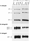
Viral protein expression in cells cotransfected with wt and mutant HXB2 proviruses. 293 cells were transfected either with 10 μg of wt HXB2 or with 5 μg each of wt and mutant HXB2 proviruses as indicated. Two days after transfection, equivalent portions of cell lysates and virion fractions were analyzed with mouse anti-p24 MAb (A), sheep anti-gp120 (B), or anti-gp41 MAb (C).

Similar articles

Cited by

References

    1. Abacioglu Y H, Fouts T R, Laman J D, Claasen E, Pincus S H, Moore J P, Roby C A, Kamin-Lewis R, Lewis G K. Epitope mapping and topology of baculovirus-expressed HIV-1 gp160 determined with a panel of murine monoclonal antibodies. AIDS Res Hum Retroviruses. 1994;10:371–381. - PubMed
    1. Bates P. Chemokine receptors and HIV-1: an attractive pair? Cell. 1996;86:1–3. - PubMed
    1. Bergeron L, Sullivan N, Sodroski J. Target cell-specific determinants of membrane fusion within the human immunodeficiency virus type 1 gp120 third variable region and gp41 amino terminus. J Virol. 1992;66:2389–2397. - PMC - PubMed
    1. Bernstein H B, Tucker S P, Kar S R, McPherson S A, McPherson D T, Dubay J W, Lebowitz J, Compans R W, Hunter E. Oligomerization of the hydrophobic heptad repeat of gp41. J Virol. 1995;69:2745–2750. - PMC - PubMed
    1. Blacklow S C, Lu M, Kim P S. A trimeric subdomain of the simian immunodeficiency virus envelope glycoprotein. Biochemistry. 1995;34:14955–14962. - PubMed

Publication types

Substances