. 1998 Jun;72(6):4832-40.
doi: 10.1128/JVI.72.6.4832-4840.1998.

Cationic liposomes enhance the rate of transduction by a recombinant retroviral vector in vitro and in vivo

Affiliations

Cationic liposomes enhance the rate of transduction by a recombinant retroviral vector in vitro and in vivo

C D Porter et al. J Virol. 1998 Jun.

Abstract

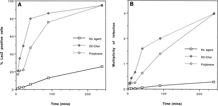
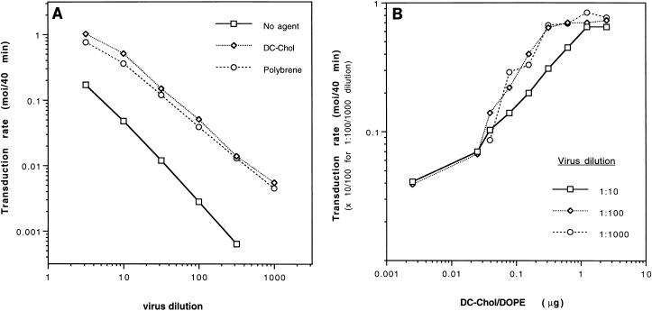
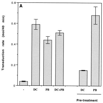
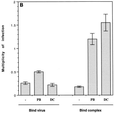
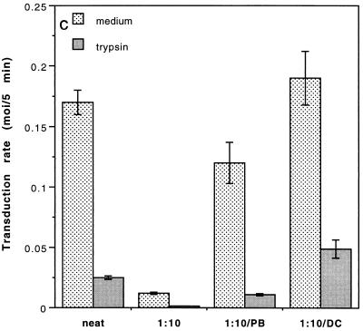
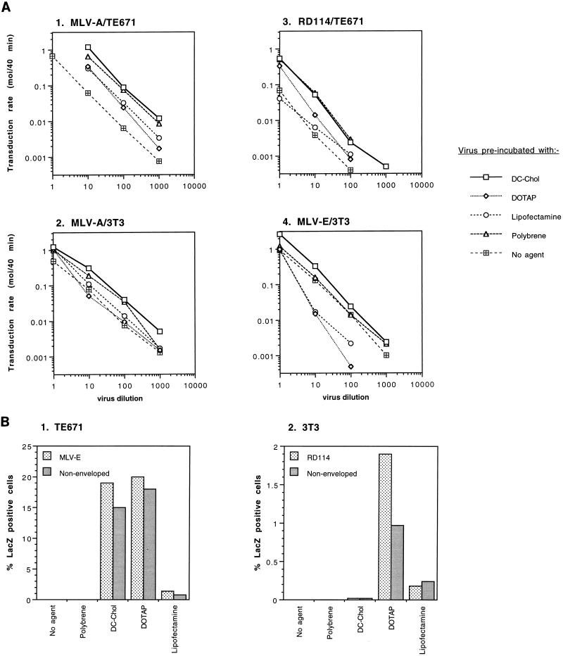
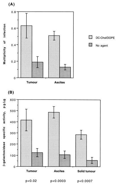
Cationic liposomes enhanced the rate of transduction of target cells with retroviral vectors. The greatest effect was seen with the formulation DC-Chol/DOPE, which gave a 20-fold increase in initial transduction rate. This allowed an efficiency of transduction after brief exposure of target cells to virus plus liposome that could be achieved only after extensive exposure to virus alone. Enhancement with DC-Chol/DOPE was optimal when stable virion-liposome complexes were preformed. The transduction rate for complexed virus, as for virus used alone or with the polycation Polybrene, showed first-order dependence on virus concentration. Cationic liposomes, but not Polybrene, were able to mediate envelope-independent transduction, but optimal efficiency required envelope-receptor interaction. When virus complexed with DC-Chol/DOPE was used to transduce human mesothelioma xenografts, transduction was enhanced four- to fivefold compared to that for virus alone. Since the efficacy of gene therapy is dependent on the number of cells modified, which is in turn dependent upon the balance between transduction and biological clearance of the vector, the ability of cationic liposomes to form stable complexes with retroviral vectors and enhance their rate of infection is likely to be important for in vivo application.


Figures

FIG. 1
FIG. 1
Transduction rate enhancement with DC-Chol/DOPE. (A) MLV-A vector (diluted 1:10) was preincubated for 30 min at room temperature with an equal volume of OptiMEM containing no agent, 50 μg of DC-Chol/DOPE (DC-Chol) per ml, or Polybrene (final concentration, 8 μg/ml) and added to TE671 target cells. After the indicated exposure time at 37°C, the reagents were replaced with medium. Two days later, histochemically lacZ-positive cells were scored as a percentage of the total cells. (B) The data in panel A were converted to MOIs enabling a more quantitative assessment of transduction efficiencies.
FIG. 2
FIG. 2
Dependence of transduction rate enhancement upon virus dilution and DC-Chol/DOPE dose. (A) Serially diluted virus was preincubated as in Fig. 1A, and target cells were exposed for 40 min to enable comparison of the initial transduction rates (MOI/40 min). (B) MLV-A vector (diluted 1:10, 1:100, or 1:1,000) was preincubated for 30 min at room temperature with serially diluted DC-Chol/DOPE (50 ng/ml to 50 μg/ml), and TE671 target cells were exposed for 40 min at 37°C to enable comparison of the initial transduction rates. No enhancement above that obtained with OptiMEM was observed with the lowest DC-Chol/DOPE dose used. For presentation purposes, the values obtained with 1:100 and 1:1,000 dilutions are multiplied by 10 and 100, respectively.
FIG. 3
FIG. 3
Order of combination and effects on virus internalization. (A) Transduction rates (MOI/40 min) were determined for preincubation of 1:10-diluted MLV-A vector with OptiMEM containing no agent (−), DC-Chol/DOPE (DC), Polybrene (PB), or a mixture of the two (DC + PB). Additionally, to evaluate the optimal order of reagent addition, target cells were pretreated with the same final concentrations of DC-Chol/DOPE (2.5 μg/350 μl) or Polybrene (8 μg/ml) for 30 min at 37°C, which was subsequently replaced with OptiMEM. Virus was added 10 min later for a 40-min exposure time. Values are mean ± SE for triplicate determinations. (B) Undiluted MLV-A vector was added to target cells on ice for 80 min (Bind virus). The cells were washed with cold OptiMEM and incubated for a further 1 h on ice in OptiMEM before being washed again to remove nonspecifically bound virus. Warm OptiMEM containing no agent (−), DC-Chol/DOPE (2.5 μg/350 μl), or polybrene (8 μg/ml) was added, and the cells were incubated for 2 h at 37°C to enable internalization of bound virus. Additionally, undiluted virus was preincubated with DC-Chol/DOPE or Polybrene for the same final concentrations and the resulting complex was cooled and added to target cells on ice for 80 min (Bind complex). After being washed as above, the cells were incubated for 2 h at 37°C in OptiMEM to enable internalization of bound virus complexes. Values are mean ± SE for triplicate determinations. (C) Undiluted (neat) and 1:10-diluted MLV-A vector, preincubated with OptiMEM containing no agent, DC-Chol/DOPE (DC), or Polybrene (PB), was added to target cells. After incubation for 5 min at 37°C, the virus was removed and replaced with medium or 0.05% trypsin and 0.02% EDTA (trypsin). Cells treated with trypsin were incubated 10 min at 37°C, after which they were recovered, washed, and replated. Values are mean ± SE for triplicate determinations.
FIG. 3
FIG. 3
Order of combination and effects on virus internalization. (A) Transduction rates (MOI/40 min) were determined for preincubation of 1:10-diluted MLV-A vector with OptiMEM containing no agent (−), DC-Chol/DOPE (DC), Polybrene (PB), or a mixture of the two (DC + PB). Additionally, to evaluate the optimal order of reagent addition, target cells were pretreated with the same final concentrations of DC-Chol/DOPE (2.5 μg/350 μl) or Polybrene (8 μg/ml) for 30 min at 37°C, which was subsequently replaced with OptiMEM. Virus was added 10 min later for a 40-min exposure time. Values are mean ± SE for triplicate determinations. (B) Undiluted MLV-A vector was added to target cells on ice for 80 min (Bind virus). The cells were washed with cold OptiMEM and incubated for a further 1 h on ice in OptiMEM before being washed again to remove nonspecifically bound virus. Warm OptiMEM containing no agent (−), DC-Chol/DOPE (2.5 μg/350 μl), or polybrene (8 μg/ml) was added, and the cells were incubated for 2 h at 37°C to enable internalization of bound virus. Additionally, undiluted virus was preincubated with DC-Chol/DOPE or Polybrene for the same final concentrations and the resulting complex was cooled and added to target cells on ice for 80 min (Bind complex). After being washed as above, the cells were incubated for 2 h at 37°C in OptiMEM to enable internalization of bound virus complexes. Values are mean ± SE for triplicate determinations. (C) Undiluted (neat) and 1:10-diluted MLV-A vector, preincubated with OptiMEM containing no agent, DC-Chol/DOPE (DC), or Polybrene (PB), was added to target cells. After incubation for 5 min at 37°C, the virus was removed and replaced with medium or 0.05% trypsin and 0.02% EDTA (trypsin). Cells treated with trypsin were incubated 10 min at 37°C, after which they were recovered, washed, and replated. Values are mean ± SE for triplicate determinations.
FIG. 4
FIG. 4
Comparison of transduction rate enhancement by different cationic liposomes for different virus-target cell combinations, and their use for envelope-independent transduction. (A) MLV-A, RD114, and MLV-E vectors were serially diluted and preincubated with OptiMEM (No agent), 50 μg of DC-Chol/DOPE (DC-Chol), DOTAP, or Lipofectamine per ml, or Polybrene (final concentration, 8 μg/ml) before exposure of TE671 or NIH 3T3 target cells for 40 min, as in Fig. 1. (B) Nonenveloped vector (derived from TELCeB6 cells) and MLV-E or RD114 vectors were preincubated with the reagents described above and used to transduce TE671 and 3T3 target cells in nonpermissive combinations by exposure for 4 h.
FIG. 5
FIG. 5
In vivo transduction efficiency of xenografted H-Meso1 cells is enhanced with DC-Chol/DOPE. MLV-A vector was preincubated with OptiMEM (No agent) or DC-Chol/DOPE and delivered intraperitoneally to nude mice bearing intraperitoneal H-Meso1 xenografts. (A) Transduction efficiencies were determined by staining cells reestablished at sacrifice in vitro from both ascites and solid tumor. (B) Lysates (five replicates for each mouse) of solid tumor deposits were used for determination of β-galactosidase specific activity. The specific activity was also determined for lysates of ascites and solid tumor cells reestablished in vitro at sacrifice. Significance levels for differences with and without liposomes are given below. All values are mean ± SE for five mice.

Similar articles

Cited by

References

    1. Aboud M, Shoor R, Salzberg S. Adsorption, penetration, and uncoating of murine leukemia virus studied by using its reverse transcriptase. J Virol. 1979;30:32–37. - PMC - PubMed
    1. Andersen K B. The fate of the surface protein gp70 during entry of retrovirus into mouse fibroblasts. Virology. 1985;142:112–120. - PubMed
    1. Andersen K B, Nexo B A. Entry of murine retrovirus into mouse fibroblasts. Virology. 1983;125:85–98. - PubMed
    1. Aubin R A, Weinfeld M, Mirzayans R, Paterson M C. Polybrene/DMSO-assisted gene transfer. Mol Biotechnol. 1994;1:29–48. - PubMed
    1. Bass D M, Baylor M R, Chen C, Mackow E M, Bremont M, Greenberg H B. Liposome-mediated transfection of intact viral particles reveals membrane penetration determines permissivity of tissue culture cells to rotavirus. J Clin Invest. 1992;90:2313–2320. - PMC - PubMed

Publication types

LinkOut - more resources