. 1998 Jun;72(6):4893-905.
doi: 10.1128/JVI.72.6.4893-4905.1998.

Long-term evolution of the hypervariable region of hepatitis C virus in a common-source-infected cohort

Affiliations

Long-term evolution of the hypervariable region of hepatitis C virus in a common-source-infected cohort

J McAllister et al. J Virol. 1998 Jun.

Abstract

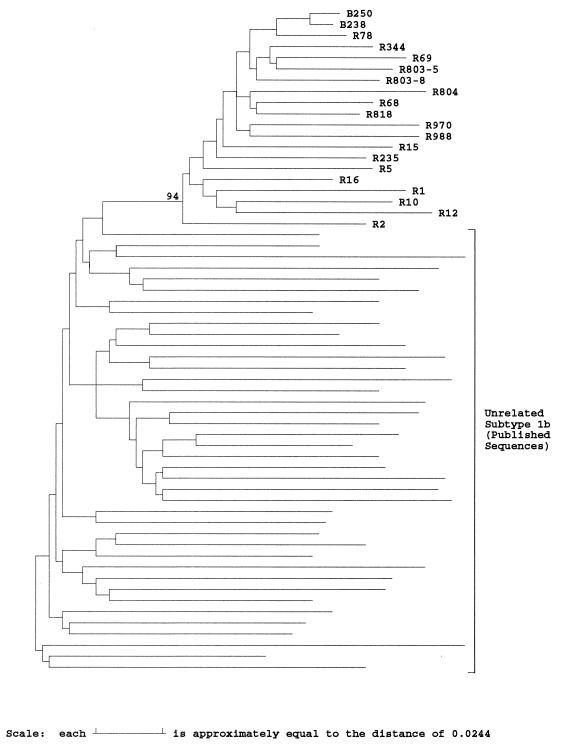
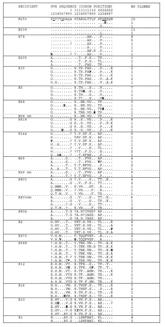
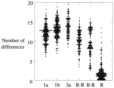
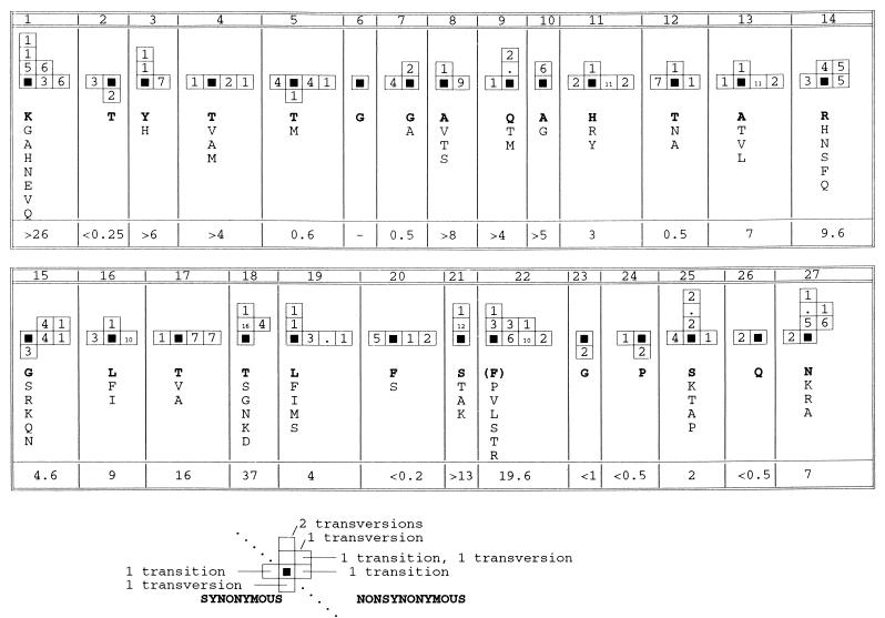
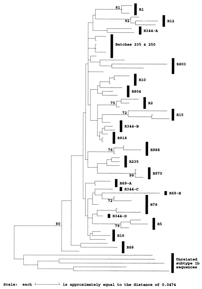
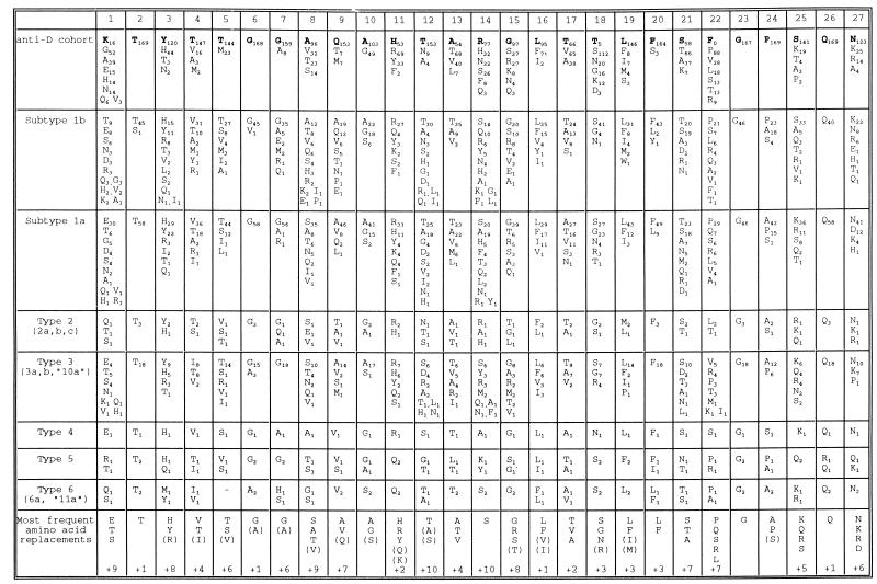
The long-term evolution of the hepatitis C virus hypervariable region (HVR) and flanking regions of the E1 and E2 envelope proteins have been studied in a cohort of women infected from a common source of anti-D immunoglobulin. Whereas virus sequences in the infectious source were relatively homogeneous, distinct HVR variants were observed in each anti-D recipient, indicating that this region can evolve in multiple directions from the same point. Where HVR variants with dissimilar sequences were present in a single individual, the frequency of synonymous substitution in the flanking regions suggested that the lineages diverged more than a decade previously. Even where a single major HVR variant was present in an infected individual, this lineage was usually several years old. Multiple lineages can therefore coexist during long periods of chronic infection without replacement. The characteristics of amino acid substitution in the HVR were not consistent with the random accumulation of mutations and imply that amino acid replacement in the HVR was strongly constrained. Another variable region of E2 centered on codon 60 shows similar constraints, while HVR2 was relatively unconstrained. Several of these features are difficult to explain if a neutralizing immune response against the HVR is the only selective force operating on E2. The impact of PCR artifacts such as nucleotide misincorporation and the shuffling of dissimilar templates is discussed.


Figures

FIG. 1
FIG. 1
Phylogenetic analysis of E1 and E2 sequences from anti-D recipients and subtype 1b isolates. Evolutionary distances between representative sequences from two anti-D immunoglobulin batches (B), 17 anti-D recipients (R), and 40 epidemiologically unrelated subtype 1b sequences in the E1/E2 region (nucleotide positions 1096 to 1458) were used to construct a neighbor-joining tree. The bootstrap support (100 replicates) for the group of sequences from anti-D recipients is indicated.
FIG. 2
FIG. 2
Phylogenetic analysis of E1/E2 sequences from anti-D recipients and infectious batches. Distinct HVR amino acid sequences (codons 1 to 27 of E2) found in cloned virus sequences from two anti-D immunoglobulin batches and 17 recipients are compared with the most frequent sequence variant present in the batches (identities shown by ., sequence ambiguities shown by ?, a stop codon shown by ∗, and single nucleotide deletions shown by #). Sequences derived from single cDNA molecules by direct sequence analysis of PCR products obtained at limiting dilution are indicated by sm. The number of clones or direct sequences that share a given HVR sequence is indicated. Amino acids underlined in the batch sequence are strongly conserved in the entire data set, while sporadic amino acid substitutions are indicated by boldface type.
FIG. 3
FIG. 3
Scatter plot of the number of amino acid differences between different groups of HVR sequences. The number of amino acid substitutions in the HVR (positions 383 to 408) was calculated for sequences of viruses of genotypes 1a (n = 58), 1b (n = 46) or 3a (n = 16), among representative sequences from different anti-D recipients (R-R), between these sequences and the consensus sequence of the infectious source (B-R), and within individual anti-D recipients (R).
FIG. 4
FIG. 4
Pattern of nucleotide substitution within the HVR. The number and type of substitutions occurring in different recipients at each codon of the HVR are summarized schematically. The solid black squares represent the nucleotide sequence of the codon found in the infectious batch at each position. Synonymous transition substitutions are represented by boxes on the horizontal axis to the left of the origin, while synonymous transversion substitutions are represented by boxes below the origin on the vertical axis. Similarly, nonsynonymous substitutions are shown to the right of the origin if they are transitions and above the origin if they are transversions. Where a codon contains multiple substitutions, this is indicated by adding the individual vectors, so that a nonsynonymous substitution produced by two transversion substitutions is indicated by two boxes on the vertical axis above the origin (see the example below the figure). The number of times each combination of substitutions was observed is indicated by the number in the corresponding box; identical substitutions occurring in different recipients were considered independent events, while sporadic substitutions were ignored. The range of amino acid residues observed at each codon is indicated, with the residue found in the infectious batch in boldface type. The ratio of the number of nonsynonymous to synonymous substitutions between the batch sequence and representative sequences from each recipient is indicated for each codon.
FIG. 5
FIG. 5
Phylogenetic analysis of the HVR flanking region at synonymous sites. Evolutionary distances between sequences at synonymous sites between positions 1096 to 1150 and 1231 to 1458 were calculated with the Jukes-Cantor correction for representative sequences from different anti-D recipients (R), two infectious batches (238 and 250), and five unrelated subtype 1b sequences and used to construct a neighbor-joining tree. Sequences from the same recipient are grouped by vertical bars, except where these group separately on the tree, in which case a suffix indicates the HVR group to which the sequences belong. Bootstrap values of 70% or more (500 replicates) are indicated.
FIG. 6
FIG. 6
Frequency of amino acid replacements within the HVR. At each position in the HVR, the frequency with which amino acid replacement is observed among different groups of sequences is indicated by a subscript. For the anti-D cohort, the amino acid present in the infectious batch is indicated by a boldface letter and sporadic substitutions are omitted. Other groups of sequences were obtained from GenBank or from published sources. The bottom row gives the most frequent amino acid replacements at each site for the whole data set, with less common replacements enclosed in parentheses and the number of additional residues observed indicated on the bottom row.

References

    1. Allander T, Beyene A, Jacobson S H, Grillner L, Persson M A A. Patients infected with the same hepatitis C virus strain display different kinetics of the isolate-specific antibody response. J Infect Dis. 1997;175:26–31. - PubMed
    1. Bean W J, Schell M, Katz J, Kawaoka Y, Naeve C, Gorman O, Webster R G. Evolution of the H3 influenza virus haemagglutinin from human and nonhuman hosts. J Virol. 1992;66:1129–1138. - PMC - PubMed
    1. Driesel G, Wirth D, Stark K, Baumgarten R, Sucker U, Schreier E. Hepatitis C virus (HCV) genotype distribution in German isolates: studies on the sequence variability in the e2 and NS5 region. Arch Virol. 1994;139:379–388. - PubMed
    1. Enomoto N, Sakuma I, Asahina Y, Kurosaki M, Murakami T, Yamamoto C, Izumi N, Marumo F, Sato C. Comparison of full-length sequences of interferon-sensitive and resistant hepatitis C virus 1b—sensitivity to interferon is conferred by amino acid substitutions in the NS5a region. J Clin Invest. 1995;96:224–230. - PMC - PubMed
    1. Esteban J I, Gomez J, Martell M, Cabot B, Quer J, Camps J, Gonzalez A, Otero T, Moya A, Esteban R, Guardia J. Transmission of hepatitis C virus by a cardiac surgeon. N Engl J Med. 1996;334:555–560. - PubMed

Publication types

Substances

Associated data

LinkOut - more resources