. 1998 Jun;72(6):5137-45.
doi: 10.1128/JVI.72.6.5137-5145.1998.

Immunoglobulin G, plasma cells, and lymphocytes in the murine vagina after vaginal or parenteral immunization with attenuated herpes simplex virus type 2

Affiliations

Immunoglobulin G, plasma cells, and lymphocytes in the murine vagina after vaginal or parenteral immunization with attenuated herpes simplex virus type 2

E L Parr et al. J Virol. 1998 Jun.

Abstract

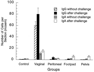
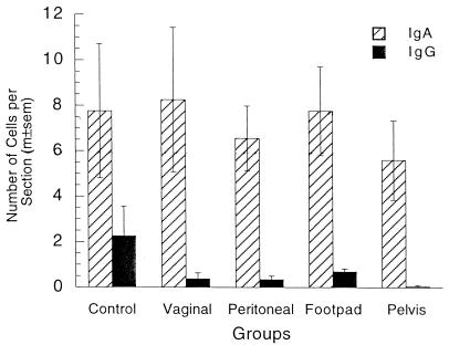
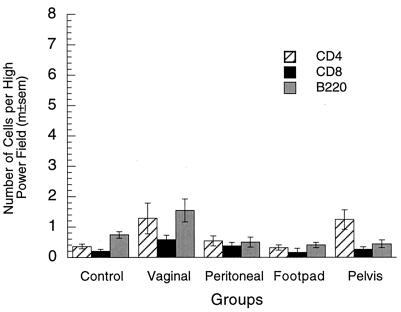
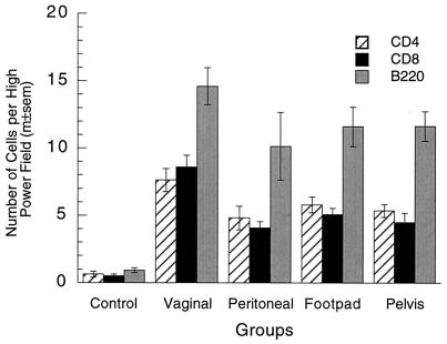
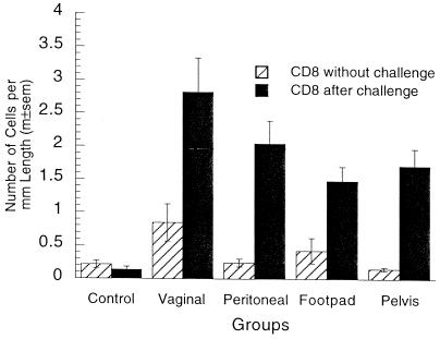
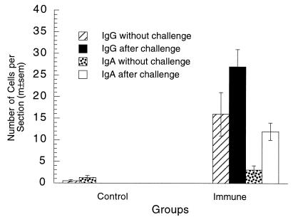
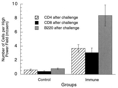
This investigation evaluated immunity to vaginal herpes simplex virus type 2 (HSV-2) infection after local or parenteral immunization with attenuated HSV-2. Vaginal immunization induced sterilizing immunity against challenge with a high dose of wild-type virus, whereas parenteral immunizations protected against neurologic disease but did not entirely prevent infection of the vagina. Vaginal immunization caused 86- and 31-fold increases in the numbers of immunoglobulin G (IgG) plasma cells in the vagina at 6 weeks and 10 months after immunization, whereas parenteral immunizations did not increase plasma cell numbers in the vagina. Vaginal secretion/serum titer ratios and specific antibody activities in vaginal secretions and serum indicated that IgG viral antibody was produced in the vagina and released into vaginal secretions at 6 weeks and 10 months after vaginal immunization but not after parenteral immunizations. In contrast to the case for plasma cells, the numbers of T and B lymphocytes in the vagina were similar in vaginally and parenterally immunized mice. Also, lymphocyte numbers in the vagina were markedly but similarly increased by vaginal challenge with HSV-2 in both vaginally and parenterally immunized mice. Lymphocyte recruitment to the vagina after virus challenge appeared to involve memory lymphocytes, because it was not observed in nonimmunized mice. Thus, local vaginal immunization with attenuated HSV-2 increased the number of IgG plasma cells in the vagina and increased vaginal secretion/serum titer ratios to 3.0- to 4.7-fold higher than in parenterally immunized groups but caused little if any selective homing of T and B lymphocytes to the vagina.


Figures

FIG. 1
FIG. 1
Plasma cells in vaginae of locally and parenterally immunized mice. The numbers of IgG and IgA plasma cells in the vagina were significantly larger in the vaginal immunization group than in the other four groups with or without vaginal challenge (P < 0.0001 in all four tests; five-group ANOVAs). Plasma cell numbers in the control and parenteral immunization groups were not significantly different without vaginal challenge (IgG, P = 0.49; IgA, P = 0.97; four-group ANOVAs). After vaginal challenge, plasma cell numbers in the parenteral immunization groups were significantly larger than those in the control group in all cases except IgG cells in the pelvic group (IgG, P = 0.0069; IgA, P = 0.0029; four-group ANOVAs), and the numbers of these cells in the parenteral immunization groups after challenge were also significantly larger than the numbers without challenge in the majority of cases (P < 0.05; two-tailed t tests). m±sem, mean ± standard error of the mean.
FIG. 2
FIG. 2
IgG plasma cells in vaginae of vaginally immunized mice. Fluorescent staining of IgG plasma cells (arrows) in a vaginal section from a mouse that was immunized in the vagina with attenuated HSV-2 6 weeks previously is shown. The cells tended to be most numerous in the periphery of the vagina and often occurred in groups. All of the cells identified as plasma cells contained cytoplasmic IgG(γ) or IgA(α). Some had the classical appearance of large ovoid cells with an eccentric nucleus, while others appeared to be smaller and to have only a thin rim of cytoplasm. It is not clear whether the latter cells were typical plasma cells in a plane of section through the nuclear side of the cell or whether they were immature plasma cells (plasmablasts). E, epithelium; L, lumen; S, stroma. Magnifications, ×121 (a) and ×315 (b).
FIG. 3
FIG. 3
Plasma cells in uteri of locally and parenterally immunized mice. Most plasma cells in the uterus contained IgA. The numbers of these cells were not significantly different in any of the groups (P = 0.90; five-group ANOVA). The number of IgG plasma cells in the uterus was significantly larger in the control group than in any of the immunized groups (P = 0.0036; five-group ANOVA). m±sem, mean ± standard error of the mean.
FIG. 4
FIG. 4
Lymphocytes in vaginae of locally and parenterally immunized mice without challenge. Lymphocyte numbers (CD4+, CD8+, and B220+) in the vaginal mucosa without challenge were somewhat larger in vaginally immunized mice than in control or parenterally immunized mice, but the statistical significance of the differences was marginal (CD4, P = 0.035; CD8, P = 0.068; B220, P = 0.0024; five-group ANOVAs). m±sem, mean ± standard error of the mean.
FIG. 5
FIG. 5
Lymphocytes in vaginae of locally and parenterally immunized mice after challenge. Vaginal lymphocyte numbers in immunized groups after vaginal challenge were 5- to 10-fold higher than those before challenge (P <0.0001 in each case; two-tailed t tests) and also 5- to 10-fold higher than those in the nonimmunized group (P < 0.0001 in all three tests; five-group ANOVAs). Lymphocyte numbers in nonimmunized mice were not significantly increased by challenge (P > 0.05 in all three tests; two-tailed t test). Lymphocyte numbers in vaginally immunized mice were somewhat larger than those in parenterally immunized mice, but the differences were not all significant (CD4, P = 0.058; CD8, P < 0.0001; B220, P = 0.29; four-group ANOVAs). m±sem, mean ± standard error of the mean.
FIG. 6
FIG. 6
CD8+ T cells in the vaginal epithelia of locally and parenterally immunized mice with and without challenge. The number of CD8+ cells in the vaginal epithelia of vaginally immunized mice without challenge was significantly larger than those in the other groups (P = 0.010; five-group ANOVA). After challenge, the numbers of CD8+ cells in the immunized groups were 3- to 10-fold larger than those before challenge (P < 0.0001 in all four tests; two-tailed t tests) and 10- to 20-fold larger than those in the nonimmunized group (P < 0.0001; five-group ANOVA). The numbers of CD8+ cells in nonimmunized mice with and without vaginal challenge were not significantly different (P = 0.30; two-tailed t test). The number of CD8+ cells after challenge was higher in vaginally immunized mice than in parenterally immunized mice, but the difference was not statistically significant (P = 0.074; four-group ANOVA). m±sem, mean ± standard error of the mean.
FIG. 7
FIG. 7
Plasma cells in vaginae of immune and control mice 10 months after vaginal immunization. Vaginal IgG and IgA plasma cell numbers 10 months after vaginal immunization were significantly larger than those in nonimmunized control mice (P < 0.0001 in each test; two-tailed t tests). These numbers were further increased after vaginal challenge. Plasma cell counts were not done in control mice after challenge. m±sem, mean ± standard error of the mean.
FIG. 8
FIG. 8
Lymphocytes in vaginae of immune and control mice 10 months after vaginal immunization. Vaginal lymphocyte numbers after vaginal challenge were 6- to 10-fold higher in mice that were immunized in the vagina 10 months previously than in nonimmune control mice (P < 0.0001 in all three tests; two-tailed t tests). m±sem, mean ± standard error of the mean.

Similar articles

Cited by

References

    1. Abraham M C, Desjardins M, Filion L G, Garber G E. Inducible immunity to Trichomonas vaginalis in a mouse model of vaginal infection. Infect Immun. 1996;64:3571–3575. - PMC - PubMed
    1. Ada G L. Vaccines. In: Paul W E, editor. Fundamental immunology. New York, N.Y: Raven Press; 1993. pp. 1309–1352.
    1. Anderson M L, BonDurant R H, Corbeil R R, Corbeil L B. Immune and inflammatory responses to reproductive tract infection with Tritrichomonas foetus in immunized and control heifers. J Parisitol. 1996;82:594–600. - PubMed
    1. Ashley R L, Corey L, Dalessio J, Wilson P, Remington M, Barnum G, Trethewey P. Protein-specific cervical antibody responses to primary genital herpes simplex virus type 2 infections. J Infect Dis. 1994;170:20–26. - PubMed
    1. Baggiolini M, Dewald B, Moser B. Interleukin-8 and related chemotactic cytokines—CXC and CC chemokines. Adv Immunol. 1994;55:97–180. - PubMed

Publication types

LinkOut - more resources