. 1998 Jun;72(6):5165-73.
doi: 10.1128/JVI.72.6.5165-5173.1998.

Complete genomic sequence of border disease virus, a pestivirus from sheep

Affiliations

Complete genomic sequence of border disease virus, a pestivirus from sheep

P Becher et al. J Virol. 1998 Jun.

Abstract

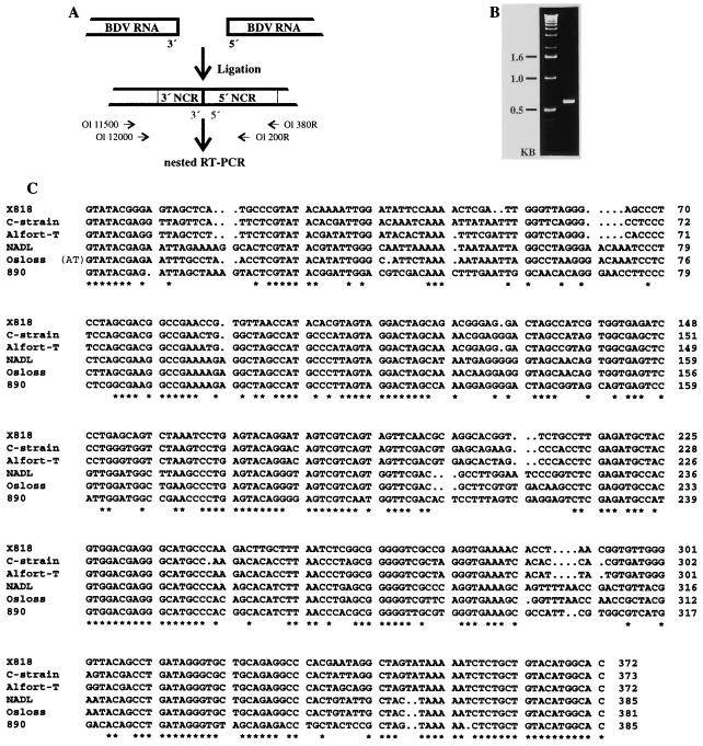
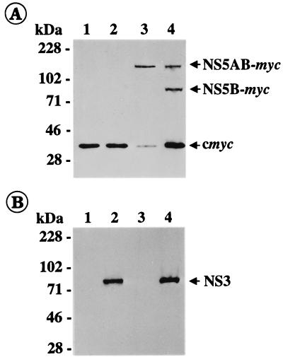
The genus Pestivirus of the family Flaviviridae comprises three established species, namely, bovine viral diarrhea virus (BVDV), classical swine fever virus (CSFV), and border disease virus from sheep (BDV). In this study, we report the first complete nucleotide sequence of BDV, that of strain X818. The genome is 12,333 nucleotides long and contains one long open reading frame encoding 3, 895 amino acids. The 5' noncoding region (NCR) of BDV X818 consists of 372 nucleotides and is thus similar in length to the 5' NCR reported for other pestiviruses. The 3' NCR of X818 is 273 nucleotides long and thereby at least 32 nucleotides longer than the 3' NCR of pestiviruses analyzed thus far. Within the 3' NCR of BDV X818, the sequence motif TATTTATTTA was identified at four locations. The same repeat was found at two or three locations within the 3' NCR of different CSFV isolates but was absent in the 3' NCR of BVDV. Analysis of five additional BDV strains showed that the 3' NCR sequences are highly conserved within this species. Comparison of the deduced amino acid sequence of X818 with the ones of other pestiviruses allowed the prediction of polyprotein cleavage sites which were conserved with regard to the structural proteins. It has been reported for two BVDV strains that cleavage at the nonstructural (NS) protein sites 3/4A, 4A/4B, 4B/5A, and 5A/5B is mediated by the NS3 serine protease and for each site a conserved leucine was found at the P1 position followed by either serine or alanine at P1' (N. Tautz, K. Elbers, D. Stoll, G. Meyers, and H.-J. Thiel, J. Virol. 71:5415-5422, 1997; J. Xu, E. Mendez, P. R. Caron, C. Lin, M. A. Murcko, M. S. Collett, and C. M. Rice, J. Virol. 71:5312-5322). Interestingly, P1' of the predicted NS5A/5B cleavage site of BDV is represented by an asparagine residue. Transient expression studies demonstrated that this unusual NS5A/5B processing site is efficiently cleaved by the NS3 serine protease of BDV.


Figures

FIG. 1
FIG. 1
5′ sequences of BDV X818. (A) Experimental strategy. After ligation of the 5′ and 3′ ends of the viral RNA, RT-PCR was performed with primers Ol 380R and Ol 11500. Two microliters of the amplification product was used for a second, nested PCR with primers Ol 200R and Ol 12000. (B) PCR product obtained after nested PCR. (C) Alignment of the 5′ NCR sequences of the four pestivirus genotypes. For BDV X818, the consensus sequence was determined from six independent clones. Other sequences were extracted from the GenBank/EMBL database (CSFV C strain [27], CSFV Alfort-T [26], BVDV-1 NADL [13], BVDV-1 Osloss [14], BVDV-2 890 [34]). Conserved nucleotides are indicated with an asterisk.
FIG. 2
FIG. 2
3′ sequences of BDV X818. (A) Experimental strategy. After ligation of an RNA oligonucleotide (Ol LIG) to the 3′ end of the viral RNA, RT-PCR was performed with primer Ol G31, which is complementary to Ol LIG, and primer Ol 11500. Two microliters of the amplification product was used for a second, seminested PCR with primers Ol G31 and Ol 11900. (B) PCR product obtained after seminested PCR. (C) Alignment of the 3′ NCR sequences of the four pestivirus genotypes. For BDV X818, the consensus sequence was determined from 12 clones obtained after ligation with an RNA oligonucleotide and from six clones obtained after ligation of the 5′ and 3′ ends of viral RNA (see also Fig. 1). Other sequences were extracted from the GenBank/EMBL database. Each sequence starts with the translational stop codon. Conserved nucleotides are indicated with an asterisk. The repeat sequence motif TATTTATTTA identified within the 3′ NCR of BDV X818, as well as CSFV C strain and Alfort-T, is underlined. (D) Predicted secondary structure of BDV X818 3′ NCR. Modeling was performed with the computer program RNAFOLD. The numbers correspond to those of the sequence shown in panel C.
FIG. 3
FIG. 3
Comparison of the 3′ NCR sequence of BDV X818 with the ones of other BDV strains. The corresponding nucleotide sequences of the 3′ NCR of ovine strains L83-84, Moredun, and Cumnock, bovine strain V-TOB, and porcine strain Frijters were determined from at least three independent clones obtained after ligation with an RNA oligonucleotide and subsequent seminested RT-PCR (for experimental strategy, see also the legend to Fig. 2). Each sequence starts with the translational stop codon. Only differences from the BDV X818 sequence are indicated. The repeat sequence motif TATTTATTTA is underlined.
FIG. 4
FIG. 4
Pestivirus polyprotein and alignment of sequences flanking the predicted cleavage sites. The diagram at the top shows the location of structural proteins (shaded boxes) and nonstructural proteins (open boxes). Proteases involved in processing include viral N-terminal autoprotease Npro, cellular signal peptidase (⧫), and viral NS2-3 (NS3) serine protease (▴). The N termini of structural proteins C, Erns, E1, and E2 have been determined for CSFV Alfort-T (37, 41), while those of the nonstructural proteins p7, NS4A, NS4B, NS5A, and NS5B have been determined for BVDV-1 strains CP7 and NADL (18, 45, 56). The alignment of sequences around the predicted processing sites include deduced amino acid sequences of CSFV Alfort-T (26), CSFV C strain (27), BVDV-1 CP7 (24), BVDV-1 NADL (13), BVDV-2 890 (34), and BDV X818 (present study).
FIG. 5
FIG. 5
Immunoblot analysis of BDV X818 nonstructural proteins. After infection with vaccinia virus MVA-T7pol, BHK-21 cells were transfected with pX818-NS34AB (lane 2), pX818-NS5AB (lane 3), or a combination of both (lane 4). BHK-21 cells infected with MVA-T7pol but not transfected served as a control (lane 1). Cells were lysed 16 h posttransfection or postinfection, and the samples were separated by sodium dodecyl sulfate-polyacrylamide gel electrophoresis (8% polyacrylamide) under reducing conditions, transferred to nitrocellulose, and incubated with anti-myc (A) or anti-NS3 (B) monoclonal antibody. The sizes of marker proteins are indicated on the left. The positions of c-myc (A) and X818-specific proteins NS5AB-myc (A), NS5B-myc (A), and NS3 (B) are marked with arrows.

References

    1. Barlow R M, Gardiner A C, Nettleton P F. The pathology of a spontaneous and experimental mucosal disease in sheep recovered from clinical border disease. J Comp Pathol. 1983;93:451–461. - PubMed
    1. Becher P, König M, Paton D, Thiel H-J. Further characterization of border disease virus isolates: evidence for the presence of more than three species within the genus pestivirus. Virology. 1995;209:200–206. - PubMed
    1. Becher P, Meyers G, Shannon A D, Thiel H-J. Cytopathogenicity of border disease virus is correlated with integration of cellular sequences into the viral genome. J Virol. 1996;70:2992–2998. - PMC - PubMed
    1. Becher P, Orlich M, Shannon A D, Horner G, Kőnig M, Thiel H-J. Phylogenetic analysis of pestiviruses from domestic and wild ruminants. J Gen Virol. 1997;78:1357–1366. - PubMed
    1. Becher P, Shannon A D, Tautz N, Thiel H-J. Molecular characterization of border disease virus, a pestivirus from sheep. Virology. 1994;198:542–551. - PubMed

Publication types

LinkOut - more resources