. 1998 Jun;72(6):5182-8.
doi: 10.1128/JVI.72.6.5182-5188.1998.

Limited transmission of Kaposi's sarcoma-associated herpesvirus in cultured cells

Affiliations

Limited transmission of Kaposi's sarcoma-associated herpesvirus in cultured cells

R Renne et al. J Virol. 1998 Jun.

Abstract

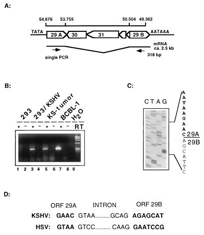
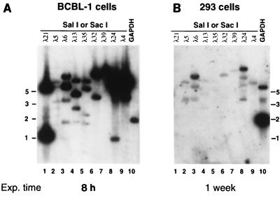
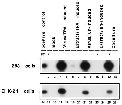
Kaposi's sarcoma-associated herpesvirus (KSHV) (also called human herpesvirus 8) is a novel gammaherpesvirus strongly implicated in the pathogenesis of Kaposi's sarcoma. Although virions can be produced in high yield from latently infected B-cell lines treated with phorbol esters, little is known about the infectivity of such virus, and efficient serial propagation of KSHV has been problematic. Here we report on the infectivity of KSHV produced from phorbol-induced BCBL-1 cells, employing an assay based on the detection of a spliced late mRNA by a sensitive reverse transcriptase PCR (RT-PCR) method. The results of this study confirm previous observations that 293 cells are susceptible to viral infection; however, infection with BCBL-1-derived virus is inefficient and the pattern of viral gene expression in infected cells may not fully reproduce that of authentic lytic infection. In keeping with this finding, serial propagation of BCBL-1-derived virus could not be demonstrated on 293 cells. Eleven of 38 other cell lines tested also supported KSHV infection, as judged by this RT-PCR assay, including cells of B-cell, endothelial, epithelial, and fibroblastic origin; however, in all cases, infection proceeded at or below the levels observed in 293 cells.


Figures

FIG. 1
FIG. 1
Mapping of the splice donor and acceptor sites of ORF29 in BCBL-1 cells and KS tumors by RT-PCR. (A) Genomic map of the region containing both exons of ORF29 divided by a 3.2-kbp region encoding four ORFs on the opposite strand. (B) Agarose gel showing the RT-PCR products. Lanes 5 and 7 show products derived from KS tumor RNA and BCBL-1 cells after 30 cycles. Lanes 1 and 2 show products derived from mock-infected or KSHV-infected 293 cells after 42 PCR cycles. As an additional control for DNA contamination, all samples were amplified in the presence (+) and absence (−) of RT. (C) After cloning of the RT-PCR product into the vector pCRII (Invitrogen), clones derived from BCBL-1 and 293 cells were sequenced and showed identical results. The sequence of the exon border between ORF29A and ORF29B is outlined. (D) Comparison of the exon and intron borders of KSHV ORF29 and herpes simplex virus (HSV) UL15; both show the conserved GT/AG major class intron consensus sequence.
FIG. 2
FIG. 2
Gene expression in BCBL-1 cells compared to that in KSHV-infected 293 cells. Phage DNA from the indicated lambda clones, which together span ca. 120 kb of the KSHV genome, was cleaved with the indicated restriction enzyme, electrophoresed through 0.8% agarose gels, stained with ethidium bromide, and transferred to duplicate nylon filters. To these filters was annealed radiolabeled cDNA probes corresponding to poly(A)+ transcripts from TPA-treated BCBL-1 cells or KSHV-infected 293 cells prepared as described in Materials and Methods. As a control for integrity of the probe as well as an index of the amount of poly(A) RNA used in the assay, a plasmid encoding glyceraldehyde phosphate dehydrogenase (GAPDH) was also electrophoresed in parallel (lane 10). Blot A was exposed for 8 h; in contrast, blot B was exposed for 1 week. Exp. time, Exposure time. (A) Hybridization of the filter with cDNA prepared from mRNA from TPA-treated BCBL-1 cells. (B) Hybridization of an identical filter with cDNA probe prepared from mRNA from KSHV-infected 293 cells. The positions of molecular size standards (in kilobases) are shown at the sides of the gels.
FIG. 3
FIG. 3
Cell-free virus is more infectious than inocula prepared from extracts. ORF29-specific RT-PCR products after 42 cycles of PCR were electrophoresed on a 0.8% agarose gel and transferred to a nylon membrane. Filters were hybridized to an ORF29-specific probe as described in Materials and Methods. As additional control for contamination, all samples were amplified in the presence (+) and absence (−) of RT. 293 and BHK-21 cells were infected with (i) cell-free virus from TPA-induced (lanes 4, 5, 17, and 18) or uninduced (lanes 8, 9, 21, and 22) BCBL-1 cells or (ii) with extracts prepared from TPA-induced (lanes 6, 7, 19, and 20) or uninduced (lanes 10, 11, 23, and 24) BCBL-1 cells. 293 and BHK-21 cells were seeded at 4 × 10⁵ cells/well in six-well plates. Monolayers were then incubated for 8 h with either cell-free virions concentrated from 10 ml of supernatant (TPA induced and uninduced) or extracts prepared from 10 ml of BCBL-1 cells at a density of 5 × 10⁵ cells/ml (TPA-induced and uninduced) as described in Materials and Methods. Monolayers were washed three times and overlaid with fresh medium, and total RNA was harvested 72 h later. In addition, 293 and BHK-21 cells were cocultivated with 106 BCBL-1 cells for 24 h (lanes 12, 13, 25, and 26). As a positive control, a diluted RT-PCR amplification product derived from BCBL-1 RNA was loaded in lanes 1 and 14.
FIG. 4
FIG. 4
KSHV isolates from four different patients behave like BCBL-1-derived virus in the ORF29-directed infectivity assay. Fresh 293 cells were incubated with 293 cell supernatants from serially passaged KSHV isolates. All supernatants which have been serially passaged up to three times scored positive in a nested DNA PCR assay (Materials and Methods). Inoculation and RT-PCR assay was done as described in the legend to Fig. 3. None of these isolates scored positive even after prolonged exposure times (lanes 1 to 10).
FIG. 5
FIG. 5
Screening for KSHV-susceptible cell lines. ORF29 RT-PCR after inoculation of cell-free virus concentrated from supernatants of TPA-induced BCBL-1 cells. A summary of all lines tested is given in Table 1. Assays were performed as described in the legend to Fig. 3. Endo, endothelial cells.

References

    1. Ambroziak J A, Blackbourn D J, Herndier B G, Glogau R G, Gullett J H, McDonald A R, Lennette E T, Levy J A. Herpes-like sequences in HIV-infected and uninfected Kaposi’s sarcoma patients. Science. 1995;268:582–583. . (Letter.) - PubMed
    1. Arvanitakis L, Mesri E A, Nador R G, Said J W, Asch A S, Knowles D M, Cesarman E. Establishment and characterization of a primary effusion (body cavity-based) lymphoma cell line (BC-3) harboring kaposi’s sarcoma-associated herpesvirus (KSHV/HHV-8) in the absence of Epstein-Barr virus. Blood. 1996;88:2648–2654. - PubMed
    1. Baines J D, Poon A P, Rovnak J, Roizman B. The herpes simplex virus 1 UL15 gene encodes two proteins and is required for cleavage of genomic viral DNA. J Virol. 1994;68:8118–8124. - PMC - PubMed
    1. Baines J D, Roizman B. The cDNA of UL15, a highly conserved herpes simplex virus 1 gene, effectively replaces the two exons of the wild-type virus. J Virol. 1992;66:5621–5626. - PMC - PubMed
    1. Blackbourn D J, Ambroziak J, Lennette E, Adams M, Ramachandran B, Levy J A. Infectious human herpesvirus 8 in a healthy North American blood donor. Lancet. 1997;349:609–611. - PubMed

Publication types

LinkOut - more resources