. 1998 Jun;72(6):5189-97.
doi: 10.1128/JVI.72.6.5189-5197.1998.

Functional interaction of human immunodeficiency virus type 1 Vpu and Gag with a novel member of the tetratricopeptide repeat protein family

Affiliations

Functional interaction of human immunodeficiency virus type 1 Vpu and Gag with a novel member of the tetratricopeptide repeat protein family

M A Callahan et al. J Virol. 1998 Jun.

Erratum in

Abstract

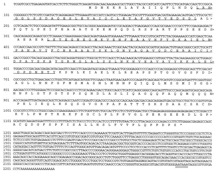
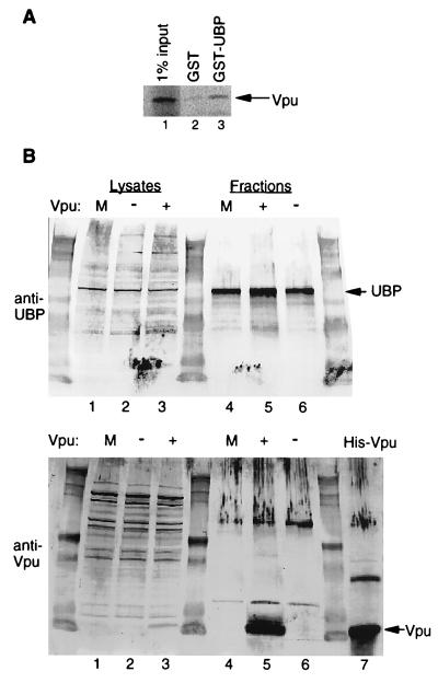
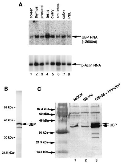
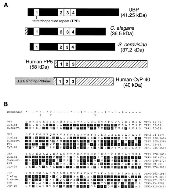
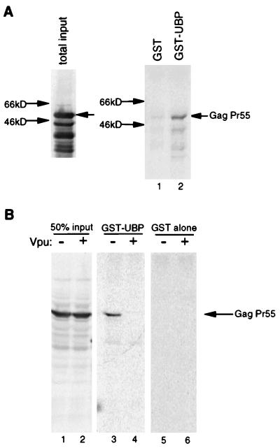
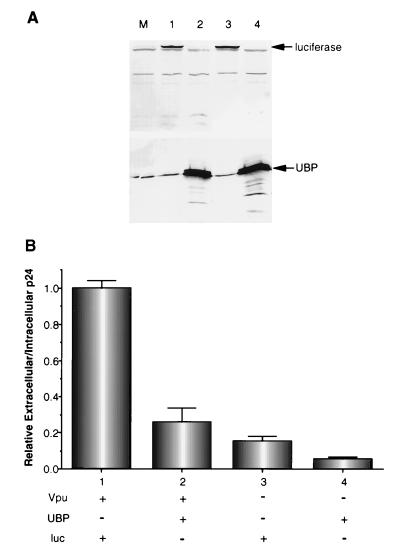
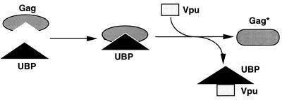
Viral protein U (Vpu) is a protein encoded by human immunodeficiency virus type 1 (HIV-1) that promotes the degradation of the virus receptor, CD4, and enhances the release of virus particles from cells. We isolated a cDNA that encodes a novel cellular protein that interacts with Vpu in vitro, in vivo, and in yeast cells. This Vpu-binding protein (UBP) has a molecular mass of 41 kDa and is expressed ubiquitously in human tissues at the RNA level. UBP is a novel member of the tetratricopeptide repeat (TPR) protein family containing four copies of the 34-amino-acid TPR motif. Other proteins that contain TPR motifs include members of the immunophilin superfamily, organelle-targeting proteins, and a protein phosphatase. UBP also interacts directly with HIV-1 Gag protein, the principal structural component of the viral capsid. However, when Vpu and Gag are coexpressed, stable interaction between UBP and Gag is diminished. Furthermore, overexpression of UBP in virus-producing cells resulted in a significant reduction in HIV-1 virion release. Taken together, these data indicate that UBP plays a role in Vpu-mediated enhancement of particle release.


Figures

FIG. 1
FIG. 1
Sequence of the ubp coding region and flanking untranslated regions. The complete nucleotide sequence of the longest ubp cDNA obtained from the two-hybrid system Vpu screen is shown with the deduced amino acid sequence of the open reading frame. Numbers to the left of each line designate nucleotide positions. The four TPR motifs are underlined, and the polyadenylation signal is double underlined.
FIG. 2
FIG. 2
Interaction between Vpu and UBP in vitro and in vivo. (A) A GST-UBP fusion protein and GST alone were expressed and purified as described in Materials and Methods. Protein concentrations were measured using the Bradford assay (Bio-Rad). Equimolar amounts of GST-UBP (lane 3) or GST alone (lane 2) were incubated in solution with 35S-labeled in vitro-translated Vpu. GST or GST-UBP was recovered on glutathione-Sepharose beads, and Vpu was detected by SDS-PAGE and phosphorimage analysis. Lane 1 shows 1% of the input Vpu used in the binding reactions. (B) Lysates of mock-transfected (m) HeLa cells (lane 1) or cells transfected with either a vpu (pBG135; lane 2) or a vpu+ (pGB108; lane 3) proviral DNA construct were subjected to anti-UBP immunoaffinity column chromatography analysis as described in Materials and Methods. Eluates from immunoaffinity columns are shown in lanes 4–6 (lane 4, mock; lane 5, pGB108; lane 6, pBG135). Cell lysates and column fractions were subjected to Western blot analysis with either anti-UBP (top) or anti-Vpu (bottom) antiserum. His-tagged E. coli-expressed Vpu is shown in lane 7.
FIG. 3
FIG. 3
(A) Northern blot analysis of ubp RNA from human cells. Top, mRNA from human spleen (lane 1), thymus (lane 2), prostate (lane 3), testis (lane 4), ovary (lane 5), small intestine (lane 6), colon (lane 7), and peripheral blood leukocytes (lane 8), probed with a ubp cDNA probe; bottom, the same blot when stripped and reprobed with a β-actin control probe. nt, nucleotides. (B) In vitro translation of UBP. The plasmid KT173 was used to express UBP in a coupled transcription-translation system (TnT; Promega) in the presence of [35S]methionine and [35S]cysteine, and protein was analyzed by SDS-PAGE and detected by phosphorimage analysis. (C) Endogenous expression and overexpression of UBP in HeLa cells. Because high level expression of UBP from pHIV-UBP is Tat dependent, plasmid pHIV-UBP was cotransfected with a plasmid that expresses HIV-1 Tat (pGB108 [16]). Lysates of mock-transfected HeLa cells (lane 1), HeLa cells transfected with pGB108 alone (lane 2), and HeLa cells transfected with pHIV-UBP and pGB108 (lane 3) were subjected to Western blot analysis using an IgG-purified rabbit polyclonal anti-UBP antibody.
FIG. 4
FIG. 4
Comparison of UBP with other members of the TPR family. (A) UBP is shown aligned with four proteins resulting from the BLAST sequence similarity search (3). TPR motifs are shown as numbered white boxes. Regions outside the TPR motifs that show sequence similarity to UBP are shown in black. Regions that do not contain sequence similarity to UBP are cross-hatched or shaded. (B) Alignment of TPR motifs in UBP and related proteins. The amino acid sequences of the TPR motifs of UBP, C. elegans (C.eleg.) R05F9.10 (accession no. U41533), S. cerevisiae (S.cerev.) UNF346 (accession no. U43491), human PP5, and human CyP-40 are shown aligned with each other and with the TPR consensus sequence. In the consensus sequence, an asterisk indicates any large hydrophobic residue, and a dash indicates any residue. Sequence identity to UBP is indicated by black reverse print. Sequence similarity to UBP is indicated by gray shading. PPIase, peptidyl-proyl cis-trans isomerase.
FIG. 5
FIG. 5
Interaction between GST-UBP and Gag expressed in bacteria and in HeLa cells. (A) Left, lysate of E. coli expressing His-Gag protein analyzed by Western blot using anti-Gag antiserum; right, result of an in vitro binding assay performed as described in Materials and Methods, using His-Gag (the total input is shown in the left-hand panel) and either GST-UBP (lane 2) or GST alone (lane 1). Gag protein from the binding assay was also detected by Western blot analysis with anti-Gag antiserum. Total protein concentrations of bacterial lysates used in the binding assay were measured by the Bradford assay. (B) Left, anti-Gag Western blot analysis of lysates of HeLa cells transfected with either a vpu (lane 1) or a vpu+ (lane 2) HIV-1 proviral construct. These lysates were used for in vitro binding assays as described in Materials and Methods, and the results are shown in the middle panel (lane 3, vpu construct; lane 4, vpu+ construct). Gag from HeLa cell lysates did not bind to GST alone (lanes 5 and 6). The presence or absence of Vpu expression in HeLa cells is indicated above the gels.
FIG. 6
FIG. 6
Effect of UBP overexpression on HIV-1 particle release. HeLa cells were mock transfected (lane M) or transfected with either pGB108 (lanes 1 and 2, Vpu+) or pBG135 (lanes 3 and 4, Vpu) and either pHIV-UBP (lanes 2 and 4, UBP+) or pTAR-luc (lanes 1 and 3, Luc+) (27). (A) Thirty-six hours posttransfection, Western blot analysis was performed on cell lysates with antigen affinity-purified antiluciferase (top) and antigen affinity-purified anti-UBP (bottom) antibodies. Lane numbers above blots correspond to numbers under the bar graph in panel B. (B) Particle release was assayed using a p24 antigen capture ELISA as described in Materials and Methods. The data are represented as the ratio of extracellular to intracellular p24 and are normalized to the GB108+TAR-luc cotransfection (bar 1). The data represent one of two independent experiments performed in triplicate. Similar results were obtained from both experiments.
FIG. 7
FIG. 7
Model of the roles of Vpu and UBP in particle release. UBP interacts with Gag forming a complex. In the absence of Vpu, this complex is stable and may be inhibitory to viral particle release. When Vpu is present, UBP is disassociated from Gag, allowing for modification(s) of Gag to form Gag*. This modification changes the protein in such a way that it can no longer interact with UBP. Either the inability of Gag* to interact with UBP or the modification of Gag itself results in enhancement of virus release.

References

    1. Adachi A, Gendelman H E, Koenig S, Folks T, Willey R, Rabson A, Martin M A. Production of acquired immunodeficiency syndrome-associated retrovirus in human and nonhuman cells transfected with an infectious molecular clone. J Virol. 1986;59:284–291. - PMC - PubMed
    1. Alkhatib G, Combadiere C, Broder C C, Feng Y, Kennedy P E, Murphy P M, Berger E A. CC CKR5: a RANTES, MIP-1alpha, MIP-1beta receptor as a fusion cofactor for macrophage-tropic HIV-1. Science. 1996;272:1955–1958. - PubMed
    1. Altschul S F, Gish W, Miller W, Myers E W, Lipman D J. Basic local alignment search tool. J Mol Biol. 1990;215:403–410. - PubMed
    1. Bour S, Schubert U, Strebel K. The human immunodeficiency virus type 1 Vpu protein specifically binds to the cytoplasmic domain of CD4: implications for the mechanism of degradation. J Virol. 1995;69:1510–1520. - PMC - PubMed
    1. Braaten D, Franke E K, Luban J. Cyclophilin A is required for an early step in the life cycle of human immunodeficiency virus type 1 before the initiation of reverse transcription. J Virol. 1996;70:3551–3560. - PMC - PubMed

Substances

LinkOut - more resources