

doi: 10.1128/JVI.72.6.5256-5261.1998.
Isolation and propagation of human papillomavirus type 16 in human xenografts implanted in the severe combined immunodeficiency mouse
Affiliations
- PMID: 9573300
- PMCID: PMC110112
- DOI: 10.1128/JVI.72.6.5256-5261.1998
Item in Clipboard
Isolation and propagation of human papillomavirus type 16 in human xenografts implanted in the severe combined immunodeficiency mouse
J Virol.
1998 Jun.
Abstract
We report the isolation and propagation of human papillomavirus type 16, the main agent of cervical cancer, using human foreskin fragments implanted in severe combined immunodeficiency mice. The infection produced viral particles, and with each passage of the virus it caused lesions identical to intraepithelial neoplasia, the precursor to carcinoma.
Figures
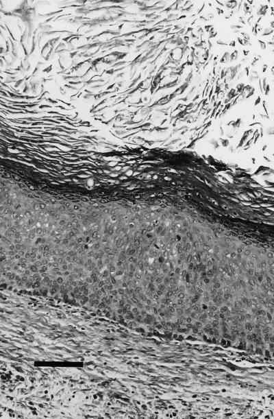
First isolation; histology (hematoxylin-eosin stain) of the single renal graft positive for the presence of HPV after the first passage. In addition to the acanthosis and parakeratosis, there is a mild basaloid proliferation and several mitotic figures are present in the stratum spinosum (bar, 600 μm).
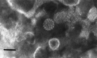
First isolation. The electron micrograph of a negatively stained preparation of the inoculum prepared from grafts collected from the second-passage experiment shows a 55-nm viral particle of morphology similar to that of papillomavirus capsids among cellular debris (bar, 55 nm).
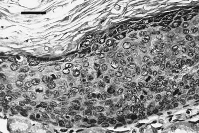
First isolation. Histology (hematoxylin-eosin stain) of one of the renal grafts from the third passage experiment shows prominent signs of intraepithelial neoplasia with basaloid proliferation, nuclear pleomorphism, dyskeratosis, and multiple aberrant mitoses throughout the stratum spinosum (bar, 100 μm).

Second isolation. A mouse from the first-passage experiment demonstrates an externalized, cutaneous graft. Magnified view of the lesion (insert) reveals a papillomatous appearance.
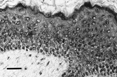
Second isolation. Histology (hematoxylin-eosin) of a subcutaneous graft from the second-passage experiment exhibits features of acanthosis, parakeratosis, and dyskeratosis (bar, 100 μm).
References
-
- Barrasso R, De Brux J, Croissant O, Orth G. High prevalence of papillomavirus-associated penile intraepithelial neoplasia in sexual partners of women with cervical intraepithelial neoplasia. N Engl J Med. 1987;317:916–923. - PubMed
-
- Bauer H M, Greer C E, Manos M M. Detection of genital human papillomavirus using PCR. In: Herrington C S, McGee J O, editors. Diagnostic molecular pathology: a practical approach. Oxford, England: Oxford University Press; 1992. pp. 131–152.
-
- Bonnez W. Papillomavirus. In: Richman D D, Whitley R J, Hayden F G, editors. Clinical virology. 1st ed. New York, N.Y: Churchill Livingstone; 1997. pp. 569–611.
-
- Bonnez, W. Unpublished data.
-
- Bonnez W, Borkhuis C, DaRin C, de Mesy Jensen K L, Beutner K, Van Nest G, Greer C. Abstracts of the 15th International Papillomavirus Workshop, Gold Coast, Queensland, Australia. 1996. Growth and propagation of human papillomavirus (HPV) type 6 in the human skin xenograft-severe combined immunodeficiency (SCID) mouse model; p. 152.
Publication types
MeSH terms
Grants and funding
LinkOut - more resources
Full Text Sources
Other Literature Sources
Medical
