

doi: 10.1128/JVI.72.6.5271-5275.1998.
Human immunodeficiency virus type 2 Vpx-Gag interaction
Affiliations
- PMID: 9573303
- PMCID: PMC110118
- DOI: 10.1128/JVI.72.6.5271-5275.1998
Item in Clipboard
Human immunodeficiency virus type 2 Vpx-Gag interaction
J Virol.
1998 Jun.
Abstract
Incorporation of Vpx into human immunodeficiency virus type 2 (HIV-2) virus-like particles is mediated by the Gag polyprotein. We have identified residues 15 to 40 of Gag p6 and residues 73 to 89 of Vpx as being necessary for virion incorporation. In addition, we show enhanced in vitro binding of Vpx to a chimeric HIV-1/HIV-2 Gag construct containing residues 2 to 49 of HIV-2 p6 and demonstrate that the presence of residues 73 to 89 of Vpx allows for in vitro binding to HIV-2 Gag.
Figures
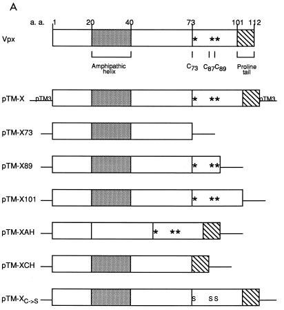
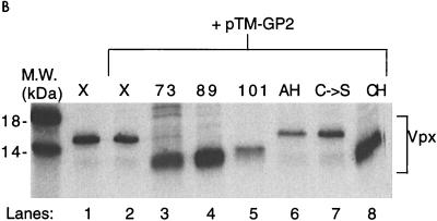
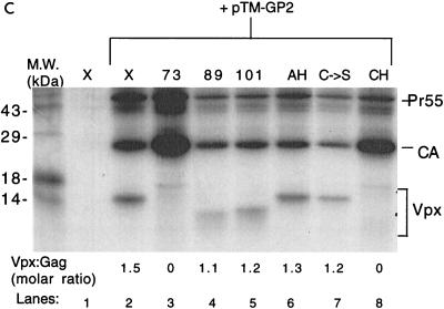
Residues 73 to 89 of HIV-2 Vpx are required for particle incorporation. (A) Schematic drawing of wild-type and mutant Vpx constructs cloned into the pTM3 vector. At the top is a diagram of HIV-2 Vpx, with the region predicted to form an amphipathic helix (shaded box), the conserved cysteines (asterisks), and the proline-rich tail (striped box) denoted. a.a., amino acids. Metabolically labeled proteins from BSC40 cell lysates (B) were immunoprecipitated with antisera to Vpx, and cell supernatants (C) were immunoprecipitated with antisera to Vpx and Gag before SDS-PAGE. The locations of the Gag and the Vpx proteins are indicated. M.W., molecular mass.
Residues 73 to 89 of HIV-2 Vpx are required for particle incorporation. (A) Schematic drawing of wild-type and mutant Vpx constructs cloned into the pTM3 vector. At the top is a diagram of HIV-2 Vpx, with the region predicted to form an amphipathic helix (shaded box), the conserved cysteines (asterisks), and the proline-rich tail (striped box) denoted. a.a., amino acids. Metabolically labeled proteins from BSC40 cell lysates (B) were immunoprecipitated with antisera to Vpx, and cell supernatants (C) were immunoprecipitated with antisera to Vpx and Gag before SDS-PAGE. The locations of the Gag and the Vpx proteins are indicated. M.W., molecular mass.
Residues 73 to 89 of HIV-2 Vpx are required for particle incorporation. (A) Schematic drawing of wild-type and mutant Vpx constructs cloned into the pTM3 vector. At the top is a diagram of HIV-2 Vpx, with the region predicted to form an amphipathic helix (shaded box), the conserved cysteines (asterisks), and the proline-rich tail (striped box) denoted. a.a., amino acids. Metabolically labeled proteins from BSC40 cell lysates (B) were immunoprecipitated with antisera to Vpx, and cell supernatants (C) were immunoprecipitated with antisera to Vpx and Gag before SDS-PAGE. The locations of the Gag and the Vpx proteins are indicated. M.W., molecular mass.
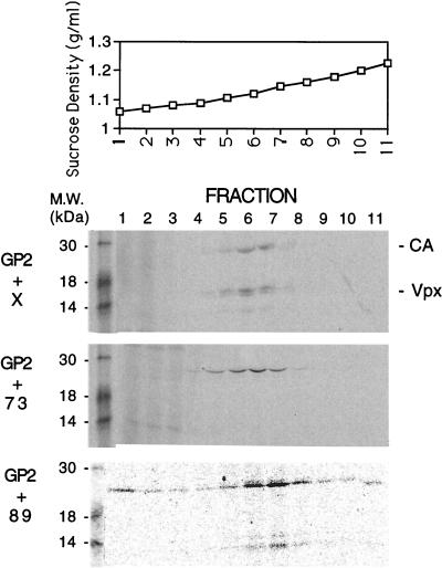
Sucrose gradient analysis of virus-like particles. The particles were concentrated by sedimentation through a 20% sucrose cushion at 26,000 rpm for 90 min in an SW28.1 rotor. Particles were resuspended in phosphate-buffered saline, layered onto linear 20 to 60% sucrose gradients, and centrifuged at 20,000 rpm for 16 h. A 20-μl aliquot of each fraction was loaded directly onto an SDS–15% PAGE gel. Fraction 1 is from the top of each gradient, and fraction 11 is from the bottom of each gradient. Protein molecular mass markers are shown in the first lane of each autoradiogram.
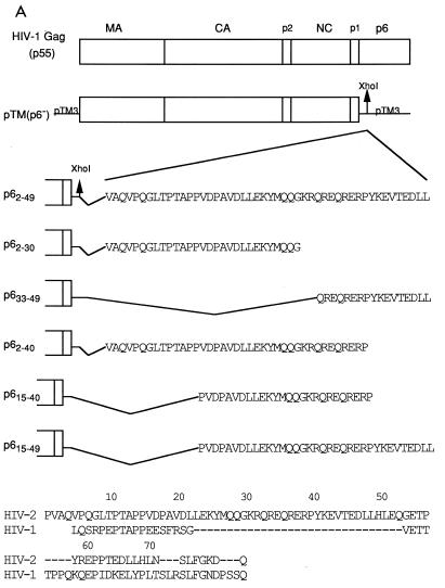
Residues 15 to 40 of HIV-2 p6 are necessary for Vpx virion incorporation. (A) Schematic drawing of the chimeric Gag constructs. Sequences of HIV-2 p6 (indicated numerically) were cloned in frame into the XhoI site of pTM(p6−). At the bottom is an alignment of HIV-2 and HIV-1 p6 sequences. (B) Cell lysates of BSC40 cells cotransfected with pTM-X and the Gag chimeric constructs were immunoprecipitated with αVpx and αGag antisera. (C) Cell supernatants from the same experiment. The locations of the Gag proteins and Vpx are indicated. M.W., molecular mass.
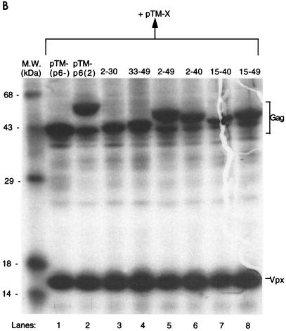
Residues 15 to 40 of HIV-2 p6 are necessary for Vpx virion incorporation. (A) Schematic drawing of the chimeric Gag constructs. Sequences of HIV-2 p6 (indicated numerically) were cloned in frame into the XhoI site of pTM(p6−). At the bottom is an alignment of HIV-2 and HIV-1 p6 sequences. (B) Cell lysates of BSC40 cells cotransfected with pTM-X and the Gag chimeric constructs were immunoprecipitated with αVpx and αGag antisera. (C) Cell supernatants from the same experiment. The locations of the Gag proteins and Vpx are indicated. M.W., molecular mass.
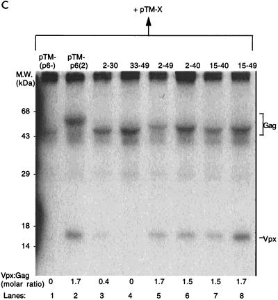
Residues 15 to 40 of HIV-2 p6 are necessary for Vpx virion incorporation. (A) Schematic drawing of the chimeric Gag constructs. Sequences of HIV-2 p6 (indicated numerically) were cloned in frame into the XhoI site of pTM(p6−). At the bottom is an alignment of HIV-2 and HIV-1 p6 sequences. (B) Cell lysates of BSC40 cells cotransfected with pTM-X and the Gag chimeric constructs were immunoprecipitated with αVpx and αGag antisera. (C) Cell supernatants from the same experiment. The locations of the Gag proteins and Vpx are indicated. M.W., molecular mass.
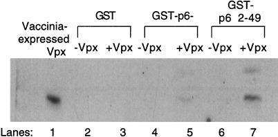
Residues 2 to 49 of HIV-2 p6 are able to enhance Vpx binding in vitro. GST and the GST-p6− and GST-p62–49 fusion proteins were incubated with vaccinia virus-expressed Vpx and glutathione beads. Proteins eluted off the beads were detected by Western blotting with Vpx antisera.
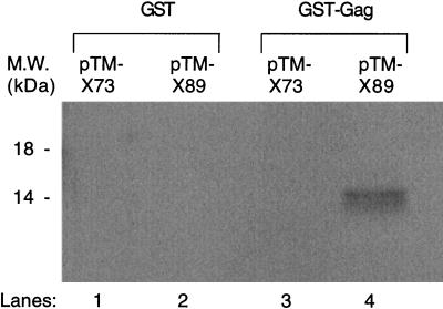
The presence of Vpx residues 73 to 89 allows for in vitro binding of Vpx to GST-Gag. GST and GST-Gag proteins were incubated with 35S-labeled Vpx73 or Vpx89 proteins. M.W., molecular mass.
Similar articles
-
Intracellular transport and virion incorporation of vpx requires interaction with other virus type-specific components.Virology. 1993 Mar;193(1):222-33. doi: 10.1006/viro.1993.1118. Virology. 1993. PMID: 8438567
-
Localization of the Vpx packaging signal within the C terminus of the human immunodeficiency virus type 2 Gag precursor protein.J Virol. 1994 Oct;68(10):6161-9. doi: 10.1128/JVI.68.10.6161-6169.1994. J Virol. 1994. PMID: 8083957 Free PMC article.
-
Vpx proteins of SIVmac239 and HIV-2ROD interact with the cytoskeletal protein alpha-actinin 1.J Gen Virol. 2004 Nov;85(Pt 11):3291-3303. doi: 10.1099/vir.0.80198-0. J Gen Virol. 2004. PMID: 15483243
-
Identification of the nuclear localization signal of human immunodeficiency virus type 2 Vpx.Virology. 2003 Jun 20;311(1):7-15. doi: 10.1016/s0042-6822(03)00093-x. Virology. 2003. PMID: 12832198
-
[Study of molecular function of proteins in human immunodeficiency virus].Yakugaku Zasshi. 2013;133(10):1103-11. doi: 10.1248/yakushi.13-00200. Yakugaku Zasshi. 2013. PMID: 24088354 Review. Japanese.
Cited by
-
Functional analysis of the simian immunodeficiency virus Vpx protein: identification of packaging determinants and a novel nuclear targeting domain.J Virol. 2001 Jan;75(1):362-74. doi: 10.1128/JVI.75.1.362-374.2001. J Virol. 2001. PMID: 11119605 Free PMC article.
-
Vif is largely absent from human immunodeficiency virus type 1 mature virions and associates mainly with viral particles containing unprocessed gag.J Virol. 2001 Jun;75(12):5504-17. doi: 10.1128/JVI.75.12.5504-5517.2001. J Virol. 2001. PMID: 11356958 Free PMC article.
-
Vpx rescues HIV-1 transduction of dendritic cells from the antiviral state established by type 1 interferon.Retrovirology. 2011 Jun 22;8:49. doi: 10.1186/1742-4690-8-49. Retrovirology. 2011. PMID: 21696578 Free PMC article.
-
A conserved dileucine-containing motif in p6(gag) governs the particle association of Vpx and Vpr of simian immunodeficiency viruses SIV(mac) and SIV(agm).J Virol. 1999 Dec;73(12):9992-9. doi: 10.1128/JVI.73.12.9992-9999.1999. J Virol. 1999. PMID: 10559313 Free PMC article.
-
In vivo infection dynamics and human adaptive changes of SIVsm-derived viral siblings SIVmac239, SIVB670 and SIVhu in humanized mice as a paralog of HIV-2 genesis.Front Virol. 2021;1:813606. doi: 10.3389/fviro.2021.813606. Epub 2021 Dec 31. Front Virol. 2021. PMID: 37168442 Free PMC article.
References
-
- Chakrabarti L, Guyader M, Alizon M, Daniel D, Desrosiers R C, Tiollais P, Sonigo P. Sequence of simian immunodeficiency virus from macaque and its relationship to other human and simian retroviruses. Nature. 1987;328:543–547. - PubMed
-
- Franchini G, Rusche J R, O’Keeffe T J, Wong-Staal F. The human immunodeficiency virus type 2 (HIV-2) contains a novel gene encoding a 16 kD protein associated with mature virions. AIDS Res Hum Retroviruses. 1988;4:243–250. - PubMed
Publication types
MeSH terms
Substances
Grants and funding
LinkOut - more resources
Full Text Sources
Other Literature Sources
Research Materials
