. 1998 Jun 1;509 ( Pt 2)(Pt 2):607-18.
doi: 10.1111/j.1469-7793.1998.607bn.x.

Paired-pulse magnetic stimulation of the human motor cortex: differences among I waves

Affiliations

Paired-pulse magnetic stimulation of the human motor cortex: differences among I waves

R Hanajima et al. J Physiol. .

Abstract

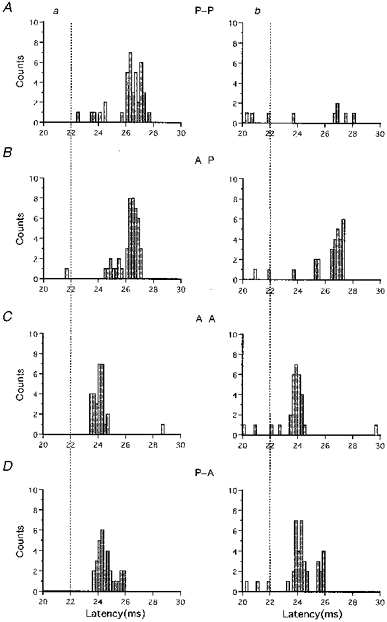
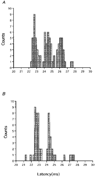
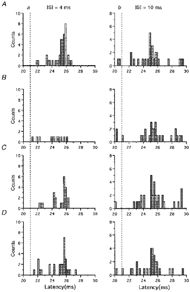
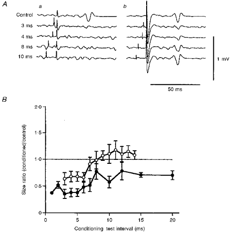
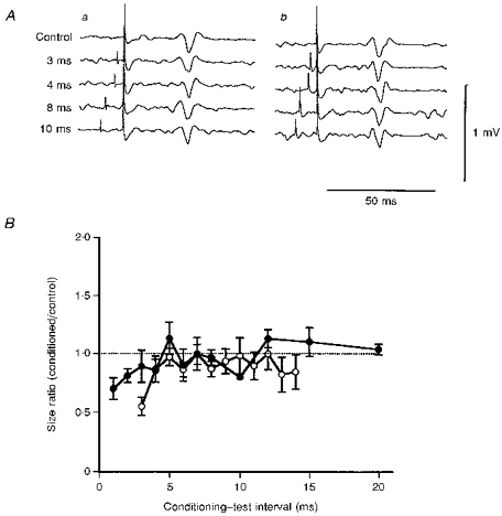
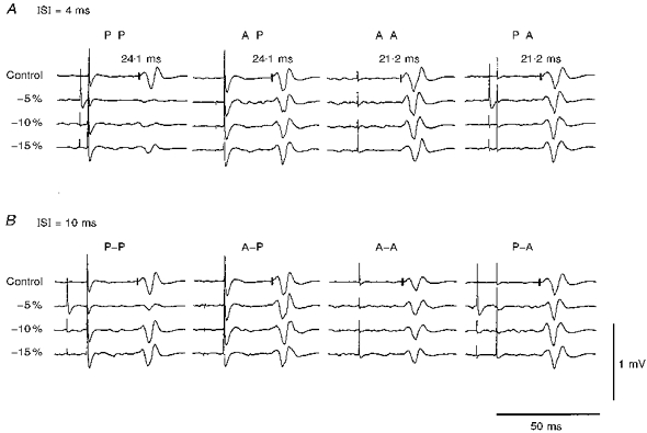
1. In paired-pulse cortical stimulation experiments, conditioning subthreshold stimuli suppress the electromyographic (EMG) responses of relaxed muscles to suprathreshold magnetic test stimuli at short interstimulus intervals (ISIs) (1-5 ms) and facilitate them at long ISIs (8-15 ms). 2. We made paired-pulse magnetic stimulation studies on the response of the first dorsal interosseous muscle (FDI) produced by I1 or I3 waves using our previously reported method which preferentially elicits one group of I waves when subjects make a slight voluntary contraction. In some experiments the conditioning and test stimuli were oppositely directed, in the others they were oriented in the same direction. Single motor unit responses were recorded with a concentric needle electrode, and surface EMG responses with cup electrodes. 3. In post-stimulus time histograms (PSTHs) of the firing probability of motor units, the peaks produced by I3 waves were decreased by a subthreshold conditioning stimulus that preferentially elicited I1 or I3 waves at an ISI of 4 ms. The amount of decrement depended on the intensity of the conditioning stimulus. The stronger the conditioning stimulus, the greater the suppression. In contrast, the peaks produced by I1 waves were little affected by any type of subthreshold conditioning stimulus, given 4 ms prior to the test stimulus. At an ISI of 10 ms, a subthreshold conditioning stimulus slightly decreased the size of the peak produced by the I3 waves, but did not affect the peaks evoked by I1 waves. 4. Surface EMGs showed that a subthreshold conditioning stimulus suppressed the responses produced by I3 waves irrespective of its current direction (anterior or posterior). Both the amount and duration of suppression depended on the intensity of the conditioning stimulus, but not on its current direction. Both parameters increased when the intensity increased. At a high intensity conditioning stimulus, suppression was evoked at ISIs of 1-20 ms, compatible with the duration of GABA-mediated inhibition found in animal experiments. Responses produced by I1 waves were little affected by any type of subthreshold conditioning stimulus. 5. We conclude that a subthreshold conditioning stimulus given over the motor cortex moderately suppresses I3 waves but does not affect I1 waves. The duration of suppression of the I3 waves supports the idea that this is an effect of GABAergic inhibition within the motor cortex.


Figures

Figure 1
Figure 1. Pairs of control and conditioned PSTHs for four different combinations of the conditioning and test stimuli in a single subject
Control PSTHs are shown on the left (a), and conditioned PSTHs at an ISI of 4 ms on the right (b). Each PSTH was constructed from 100 trials. The abscissa shows the latency after the magnetic stimulation for 20-30 ms. A, P-P is the condition under which both the conditioning and test stimuli were posteriorly directed current in the brain. B, in A-P, the conditioning stimulus was anteriorly directed current, and the test stimulus was posteriorly directed current. C, in A-A, both stimuli were anteriorly directed current. D, in P-A, the conditioning stimulus was posteriorly directed current, and the test stimulus anteriorly directed. In the control PSTHs (Aa and Ba) the posteriorly directed test stimulus elicited a single peak with a latency about 4.5 ms later than that of the peak produced by electrical stimulation (D wave, dotted lines). The peak was much reduced by the conditioning stimulus under the P-P condition (Ab), and moderately so under the A-P condition (Bb). In the control PSTHs, the anteriorly directed test stimulus produced a single peak 2 ms later than the D wave (Ca and Da). These peaks were not reduced by both the anteriorly and posteriorly directed conditioning stimuli (Cb and Db).
Figure 2
Figure 2. Effect of a conditioning stimulus on three peaks in a PSTH (total 100 trials) evoked by an anteriorly directed test stimulus of high intensity
A, the anteriorly directed test stimulus of high intensity produced three peaks in a PSTH which had latencies compatible with I1, I2 and I3 waves. B, the anteriorly directed conditioning stimulus of -5 % intensity given at an ISI of 4 ms markedly decreased the size of the I3 wave, moderately decreased that of the I2 wave, but did not affect the I1 wave.
Figure 3
Figure 3. Dependence of inhibition on the intensity of the conditioning stimulus under the P-P condition
A shows the control PSTHs; B-D show conditioned PSTHs. Total trial number was 100 for every PSTH. Conditioned PSTHs at an ISI of 4 ms are given on the left (a) and those at an ISI of 10 ms on the right (b). The test stimulus evoked a peak that corresponded to the I3 wave. Conditioning stimuli of -5, -10 and -15 % intensity reduced the peak size produced by the test stimulus at an ISI of 4 ms. In the PSTH for the -5 % conditioning stimulus given 10 ms prior to the test stimulus, the peak produced by the test stimulus was smaller than that in the control PSTH. This peak was not affected by conditioning stimuli of -10 or -15 % intensity given at an ISI of 10 ms.
Figure 4
Figure 4. Averaged surface EMG potentials (A) and mean (±s.e.m.) time courses (B) for responses evoked by a posteriorly directed test stimulus
Aa, responses under the P-P condition; Ab, responses under the A-P condition. Top traces show the control responses and the four lower traces show responses to a test stimulus preceded by a conditioning stimulus by 3, 4, 8 and 10 ms. The conditioning stimulus was fixed at 5 % less than the threshold for the active muscle. The sizes of control responses were about 0.2 mV. Conditioned responses for the P-P condition were significantly smaller than the control responses at all the ISIs (Student's unpaired t test: P < 0.01). Those for the A-P condition were significantly smaller than the control at ISIs of 3 and 4 ms (Student's unpaired t test: P < 0.02), but not significantly different at ISIs of 8 and 10 ms (P > 0.2). B, mean (±s.e.m.) time courses of the effect on the response to a posteriorly directed test stimulus were obtained from the results for all the subjects. •, P-P condition; ○, A-P condition. Each point represents the mean (±s.e.m.) size ratio for all the subjects. The abscissa shows the ISIs and the ordinate the size ratios. In both time courses, significant suppression occurred (ANOVA test: P < 0.01 for P-P, P < 0.02 for P-A). At short ISIs (1-5 ms), the size ratios for both conditions were significantly less than 1.0 (Tukey's method: P < 0.01 for P-P, P < 0.05 for A-P). Significant suppression continued up to an ISI of 20 ms in the time course for the P-P condition (P < 0.05 for ISI of 20 ms), but not in that for the A-P condition. Significantly deeper suppression occurred under the P-P condition than under the A-P condition (ANOVA test: P < 0.05)
Figure 5
Figure 5. Averaged surface EMG responses (A) and mean (±s.e.m.) time courses (B) for the A-A and P-A conditions
The arrangement is the same as in Fig. 4. Aa, responses under the A-A condition; Ab, responses under the P-A condition. The intensity of the conditioning stimulus was -5 %. The conditioned response was smaller than the control response only at an ISI of 3 ms under the A-A condition (Aa). This suppression was not significant (Student's unpaired t test: P > 0.1). No suppression occurred at the other intervals under the A-A condition (Aa). No suppression was evoked at any intervals under the P-A condition (Ab). B, slight but significant suppression occurred at ISIs of 1 and 2 ms in the time course for the A-A condition (•; Student's unpaired t test: P < 0.05), and only at an ISI of 3 ms for the P-A condition (○; Student's unpaired t test: P < 0.05).
Figure 6
Figure 6. Typical responses of a single subject to a given test stimulus conditioned by stimuli of different intensities
Averaged control and conditioned responses at ISIs of 4 ms (A) and 10 ms (B) are shown. The left column shows responses for the P-P condition, and the other columns responses for the A-P, A-A and P-A conditions. The conditioning stimulus intensities were 5, 10 and 15 % less than the threshold for the active muscles. All the control responses were similar in size. A, significant suppression occurred under the P-P conditions at all the intensities used (Student's unpaired t test: P < 0.01) and under the A-P condition with a -5 % conditioning stimulus given at an ISI of 4 ms (Student's unpaired t test: P < 0.05).B, at an ISI of 10 ms, however, there was significant suppression only for a -5 % conditioning stimulus under the P-P condition (Student' s unpaired t test: P < 0.01).

References

    1. Bekenstein J, Rempe D, Lothman E. Decreased heterosynaptic and homosynaptic paired-pulse inhibition in the rat hippocampus as a chronic sequel to limbic status epilepticus. Brain Research. 1993;601:111–120. - PubMed
    1. Hanajima R, Ugawa Y, Terao Y, Ogata K, Kanazawa I. Ipsilateral cortico-cortical inhibition of the motor cortex in various neurological disorders. Journal of the Neurological Sciences. 1996;140:109–116. 10.1016/0022-510X(96)00100-1. - DOI - PubMed
    1. Krnjevic K, Randic M, Straughan DW. Cortical inhibition. Nature. 1964;201:1294–1296. - PubMed
    1. Krnjevic K, Randic M, Straughan DW. Pharmacology of cortical inhibition. The Journal of Physiology. 1965;184:78–105. - PMC - PubMed
    1. Kujirai T, Caramia MD, Rothwell JC, Day BL, Thompson PD, Ferbert A, Wroe S, Asselman P, Marsden CD. Cortico-cortical inhibition in human motor cortex. The Journal of Physiology. 1993;471:501–519. - PMC - PubMed

Publication types

LinkOut - more resources