. 1998 May;117(1):43-53.
doi: 10.1104/pp.117.1.43.

Expression pattern of the carrot EP3 endochitinase genes in suspension cultures and in developing seeds

Affiliations

Expression pattern of the carrot EP3 endochitinase genes in suspension cultures and in developing seeds

van Hengel AJ et al. Plant Physiol. 1998 May.

Abstract

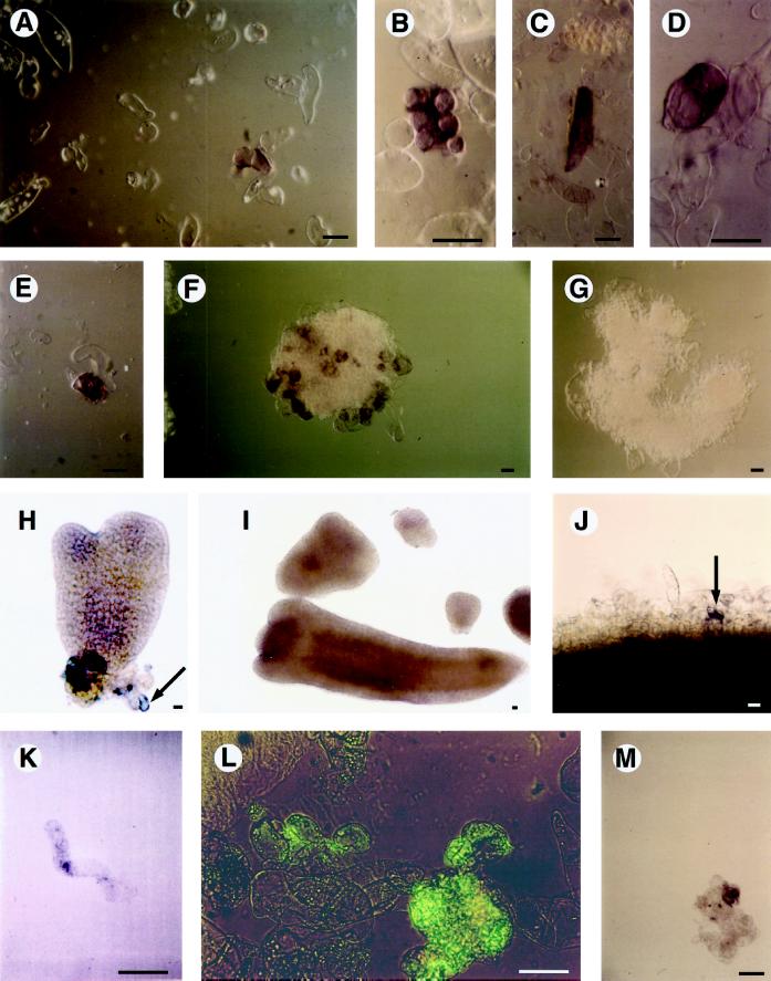
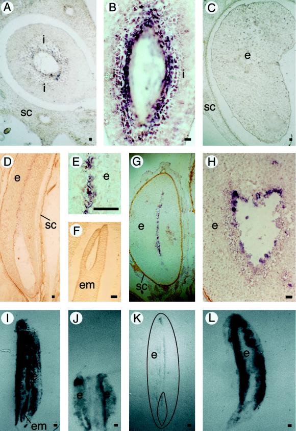
Carrot (Daucus carota) extracellular protein 3 (EP3) class IV endochitinases were previously identified based on their ability to rescue somatic embryos of the temperature-sensitive cell line ts11. Whole-mount in situ hybridization revealed that a subset of the morphologically distinguishable cell types in embryogenic and nonembryogenic suspension cultures, including ts11, express EP3 genes. No expression was found in somatic embryos. In carrot plants EP3 genes are expressed in the inner integumentary cells of young fruits and in a specific subset of cells located in the middle of the endosperm of mature seeds. No expression was found in zygotic embryos. These results support the hypothesis that the EP3 endochitinase has a "nursing" function during zygotic embryogenesis and that this function can be mimicked by suspension cells during somatic embryogenesis.


Figures

Figure 1
Figure 1
EP3 gene expression in suspension cultures. A to D, Examples of EP3-expressing cells in an embryogenic cell culture. E and F, Cell clusters present in an embryogenic cell culture. G, Cell cluster present in an embryogenic cell culture hybridized with an EP3 sense probe. H, Heart-stage somatic embryo; the arrow points to an attached EP3-expressing single cell. I, Torpedo-stage somatic embryo devoid of EP3 expression. J, Cells present at the periphery of the proliferated cell mass on a section of a hypocotyl explant that was cultured in the presence of 2,4-D for 10 d; the arrow points to a single Ep3-expressing cell. K, An elongated single cell released from a hypocotyl explant that was cultured in the presence of 2,4-D for 20 d. L, Cell clusters present in an embryogenic cell suspension subjected to immersion immunofluorescence; the presence of the EP3 protein is indicated by the green fluorescence. M, Cell cluster in a suspension of the mutant cell line ts11; the presence of the EP3 protein is indicated by a purple precipitate. Plant material was analyzed by whole-mount in situ hybridization with antisense EP3 RNA probes (A–F and H–K) or with a sense EP3 RNA probe (G). Light microscopy (coupled with Nomarski optics for A–E and M) was used for visualization of the purple precipitate (the use of Nomarski optics resulted in a change from purple to brown in A–F and J). In H, EP3 gene expression is visible as a purple precipitate in an individual cell. Immunolocalization of EP3 proteins in suspension cultures (L and M) was done by immersion immunofluorescence using either fluorescein isothiocyanate (L) or alkaline phosphatase as a second antibody. Bars = 50 μm. {/ANNT;152064n;3872n;center;704n}
Figure 2
Figure 2
Presence of EP3 chitinases in media of cultures treated with fungi and elicitors. Media of suspension cultures grown in the presence of: lane 1, 0.002 mg/mL chitosan; lane 2, 0.02 mg/mL chitosan; lane 3, P. infestans; lane 4, B. cinerea; and lane 5, control suspension culture. Proteins were separated by denaturing SDS-PAGE and detected by immunoblotting.
Figure 3
Figure 3
Presence of EP3 chitinases in media of nonembryogenic, embryogenic, and density-fractionated embryogenic cultures. A, Detection of EP3 isoenzymes in the medium of several different suspension cultures. Lanes 1 and 2, Nonembryogenic suspension cultures; and lanes 3 to 6, embryogenic suspension cultures. B, Detection of EP3 isoenzymes in the medium of a Percoll-fractionated suspension culture: lane 1, cells < 50 μm; lane 2, 0 to 10% Percoll; lane 3, 10 to 20% Percoll; lane 4, 20 to 30% Percoll; lane 5, 30 to 40% Percoll; and lane 6, cells > 125 μm. The proteins were separated by nondenaturing PAGE and detected by immunoblotting.
Figure 4
Figure 4
Expression of EP3 genes in carrot plants in the leaf (lane 1), stem (lane 2), storage root (lane 3), root (lane 4), flower (lane 5), fruit harvested 3 DAP (lane 6), fruit harvested 7 DAP (lane 7), fruit harvested 10 DAP (lane 8), fruit harvested 20 DAP (lane 9), mature seed (lane 10), seed 12 h after imbibition (lane 11), seed 60 h after imbibition (lane 12), genomic DNA (lane 13), and water control (lane 14). B, Ubiquitin control. Gene expression was determined by RT-PCR, followed by transfer to membranes and Southern hybridizaton.
Figure 5
Figure 5
EP3 gene expression in seeds. A, Cross-section of a fruit 7 DAP. B, Longitudinal section of a fruit 7 DAP. C, Cross-section of a fruit 20 DAP. D, Longitudinal section of a mature seed. E, Longitudinal section of a mature seed. F, Zygotic embryo in a longitudinal section of a mature seed. G, Transverse section of a seed that soaked in water for 60 h. H, Transverse section of a seed that soaked in water for 60 h. In the immunolocalizations, the presence of EP3 proteins is visible as a dark precipitate. Bar = 100 μm. I, Control tissue print of a mature seed stained with amido black. J, Immunolocalization of EP3 on a tissue print of a fruit containing two developing seeds 20 DAP. K, Immunolocalization of EP3 on a tissue print of a mature seed (a drawing of the printed seed is superimposed on the picture). L, Immunolocalization of EP3 on a tissue print of a seed that soaked in water for 60 h. Plant material was analyzed by in situ hybridization on sectioned carrot seeds with antisense EP3 RNA probes (A–H). Light microscopy (coupled to Nomarski optics for A–E) was used for visualization of the purple precipitate (the use of Nomarski optics resulted in a change from purple to brown in D). Immunolocalization of the EP3 protein was done by tissue printing followed by immunostaining (I–L). i, Integuments surrounding the developing embryo and endosperm; SC, seed coat; e, endosperm; em, embryo. Bar = 50 μm.
Figure 6
Figure 6
Comparison of EP3 chitinases in suspension culture and in seeds. Lane 1, Proteins obtained from the conditioned medium of a suspension culture; and lane 2, proteins obtained from mature, dry carrot seeds. The proteins were separated by nondenaturing PAGE, and the EP3 chitinases were detected by immunoblotting.

Similar articles

Cited by

References

    1. Bewley JD, Black M (1994) Seeds: Physiology of Development and Germination, Ed 2. Plenum Press, New York
    1. Borthwick HA. Development of the macrogametophyte and embryo of Daucus carota. Bot Gaz. 1931;92:23–44.
    1. Bulawa CE, Wasco W. Chitin and nodulation. Nature. 1991;353:710. - PubMed
    1. Cox KH, Goldberg RB. Analysis of plant gene expression. In: Shaw CH, editor. Plant Molecular Biology: A Practical Approach. Oxford, UK: IRL Press; 1988. pp. 1–34.
    1. De Jong AJ, Cordewener J, Lo Schiavo F, Terzi M, Vandekerckhove J, Van Kammen A, De Vries SC. A carrot somatic embryo mutant is rescued by chitinase. Plant Cell. 1992;4:425–433. - PMC - PubMed

LinkOut - more resources