. 1998 May;117(1):73-84.
doi: 10.1104/pp.117.1.73.

A common position-dependent mechanism controls cell-type patterning and GLABRA2 regulation in the root and hypocotyl epidermis of Arabidopsis

Affiliations

A common position-dependent mechanism controls cell-type patterning and GLABRA2 regulation in the root and hypocotyl epidermis of Arabidopsis

C Y Hung et al. Plant Physiol. 1998 May.

Abstract

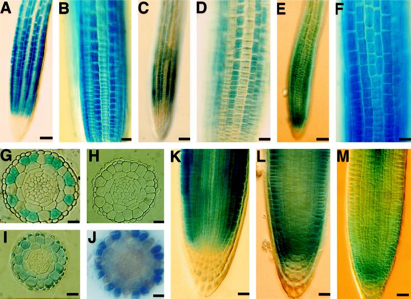
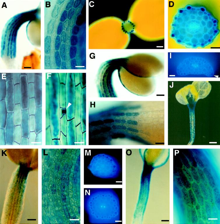
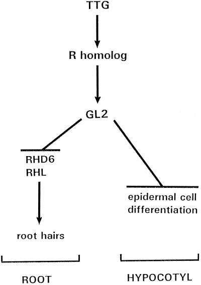
A position-dependent pattern of epidermal cell types is produced during root development in Arabidopsis thaliana. This pattern is reflected in the expression pattern of GLABRA2 (GL2), a homeobox gene that regulates cell differentiation in the root epidermis. GL2 promoter::GUS fusions were used to show that the TTG gene, a regulator of root epidermis development, is necessary for maximal GL2 activity but is not required for the pattern of GL2 expression. Furthermore, GL2-promoter activity is influenced by expression of the myc-like maize R gene (35S::R) in Arabidopsis but is not affected by gl2 mutations. A position-dependent pattern of cell differentiation and GL2-promoter activity was also discovered in the hypocotyl epidermis that was analogous to the pattern in the root. Non-GL2-expressing cell files in the hypocotyl epidermis located outside anticlinal cortical cell walls exhibit reduced cell length and form stomata. Like the root, the hypocotyl GL2 activity was shown to be influenced by ttg and 35S::R but not by gl2. The parallel pattern of cell differentiation in the root and hypocotyl indicates that TTG and GL2 participate in a common position-dependent mechanism to control cell-type patterning throughout the apical-basal axis of the Arabidopsis seedling.


Figures

Figure 1
Figure 1
Effect of GL2-promoter fragments on GUS-reporter expression in roots and hypocotyls of Arabidopsis seedlings. The GL2-promoter fragments fused to the GUS-reporter gene are shown on the left. The ability of these constructs to drive GUS expression in a cell-position-dependent manner in the seedling root and/or hypocotyl was determined by histochemical staining with the X-Gluc substrate. +, Typical GUS expression pattern detected; −, abnormal/no GUS expression detected. The relative root/hypocotyl GUS-activity value was determined by comparing the GUS activity (calculated as millimoles of product per milligram of protein per minute) in root versus hypocotyl extracts from a common set of 3-d-old seedlings bearing the indicated transgene. Values are means ± sd. Xh, XhoI; H, HindIII; Hp, HpaI; RI, EcoRI; RV, EcoRV; X, XbaI; M, MscI; and ▵, deletion.
Figure 2
Figure 2
Spatial expression pattern of GL2::GUS-reporter-gene fusion construct during root development in Arabidopsis seedlings. Four-day-old seedlings were assayed for GUS activity by histochemical staining with the X-Gluc substrate. A, Wild-type root containing the GL2::GUS transgene. Bar = 50 μm. B, Wild-type root containing the GL2::GUS transgene. GUS-expressing cells are preferentially located in specific epidermal cell files. Bar = 20 μm. C, ttg-1 mutant root containing the GL2::GUS transgene. Bar = 50 μm. D, ttg-1 mutant root containing the GL2::GUS transgene. GUS-expressing cells are preferentially located in specific epidermal cell files. Bar = 20 μm. E, 35S::R root containing the GL2::GUS transgene. Bar = 50 μm. F, 35S::R root apex containing the GL2::GUS transgene. GUS-expressing cells are not clearly located in specific epidermal cell files. Bar = 20 μm. G, Wild-type root containing the GL2::GUS transgene; transverse plastic section taken from the late meristematic region. GUS expression is limited to epidermal cells located outside periclinal cortical cell walls (i.e. in contact with a single cortical cell). At this developmental stage, a single layer of lateral root cap cells surrounds the epidermis. Bar = 20 μm. H, ttg-1 mutant root containing the GL2::GUS transgene; transverse plastic section taken from the late meristematic region. No GUS expression is observed. At this developmental stage, a single layer of lateral root cap cells surrounds the epidermis. Bar = 20 μm. I, 35S::R root containing the GL2::GUS transgene; transverse plastic section taken from the late meristematic region. GUS expression is observed throughout the epidermis, cortex, and lateral root cap. At this developmental stage, a single layer of lateral root cap cells surrounds the epidermis. Bar = 20 μm. J, ttg-1 mutant root containing the GL2::GUS transgene; thick transverse section from agarose-embedded root. GUS expression is observed in epidermal cells located outside periclinal cortical cell walls. Bar = 20 μm. K, Wild-type root apex containing the GL2::GUS transgene. Whole-mount root preparation showing GUS expression near the meristem initials but not within the lateral or columella root cap cells. The dense staining visible in the upper portion of this root is due to GUS-expressing epidermal cells above and below the plane of focus. Bar = 20 μm. L, 35S::R root apex containing the GL2::GUS transgene. Whole-mount root preparation showing GUS expression throughout region containing meristem initials and within the lateral root cap cells but not within the columella root cap cells. The dense staining visible in the upper root is due to GUS-expressing cells above and below the plane of focus. Bar = 20 μm. M, 35S::GUS root apex. Whole-mount root preparation showing preferential GUS expression throughout region containing meristem initials, the root cap, and the developing vascular tissue. Bar = 20 μm. These GL2::GUS seedlings all contain the full-length 4-kb GL2 promoter::GUS transgene. Note that root hairs are not visible in these images near the root apex; hairs form on epidermal cells at a later developmental age, just beyond the field of view shown in A, C, and E.
Figure 3
Figure 3
The spatial-expression pattern of the GL2-GUS-reporter-gene fusion construct during hypocotyl development. Seedlings harboring the 4-kb GL2 promoter::GUS transgene were stained for GUS activity using X-Gluc. A, Wild-type hypocotyl from 3-d-old seedling. Bar = 100 μm. B, Wild-type hypocotyl epidermis from 3-d-old seedling. GUS-expressing cells are located within specific epidermal cell files. Bar = 50 μm. C, Wild-type 3-d-old seedling sectioned through the hypocotyl. A ring of GUS-expressing hypocotyl epidermal cells is visible. Bar = 100 μm. D, Wild-type hypocotyl from 3-d-old seedling; transverse agarose section. GUS expression is present in epidermal cells located outside a periclinal cortical cell wall. E, Wild-type hypocotyl from 5-d-old seedling. Note that cells in the GL2::GUS-expressing files are longer than cells in the nonexpressing files. Bar = 40 μm. F, Wild-type hypocotyl from 5-d-old seedling. Stomatal development (arrowhead) occurs in non-GL2::GUS-expressing cell files. Bar = 40 μm. G, Wild-type hypocotyl and cotyledons from 3-d-old seedling. GUS-expressing cells are visible at the margin of cotyledons. Bar = 100 μm. H, Wild-type hypocotyl/cotyledon junction region from 3-d-old seedling. Bar = 50 μm. I, Wild-type cotyledon; transverse section taken near cotyledon apex (3-d-old). Arrowhead indicates a GUS-staining epidermal cell. Bar = 50 μm. J, Wild-type hypocotyl from 7-d-old seedling. GUS expression is visible in the developing leaf primordia and trichomes. Bar = 200 μm. K, ttg-1 mutant hypocotyl from 3-d-old seedling. Bar = 100 μm. L, ttg-1 mutant hypocotyl from 3-d-old seedling. GUS-expressing cells appear to be located within specific epidermal cell files. Bar = 50 μm. M, ttg-1 mutant hypocotyl from 3-d-old seedling; transverse agarose section. GUS expression is present in epidermal cells located outside a periclinal cortical cell wall (i.e. in contact with a single cortical cell). Bar = 50 μm. N, 35S::R hypocotyl from 3-d-old seedling; transverse agarose section. GUS expression is present in cells located throughout the epidermis. Bar = 50 μm. O, 35S::R hypocotyl from 3-d-old seedling. Bar = 100 μm. P, 35S::R hypocotyl from 3-d-old seedling. GUS-expressing cells are located throughout the epidermis. Bar = 50 μm.
Figure 4
Figure 4
Proposed pathway for the regulation of cell differentiation in the root and hypocotyl of Arabidopsis. The TTG is proposed to activate an R-like bHLH protein that positively controls the transcription of GL2. The GL2 homeodomain protein is proposed to control root and hypocotyl epidermal cell differentiation. See text for additional discussion. Arrows indicate positive action; blunted lines indicate negative regulation. RHD6, Root hair defective 6; and RHL, root hairless.

Similar articles

Cited by

References

    1. Avers CJ. Fine structure of phleum root meristem cells. II. Mitotic asymmetry and cellular differentiation. Am J Bot. 1963;50:140–148.
    1. Berger F, Hung C-Y, Dolan L, Schiefelbein J (1998) Control of epidermal cell division in the root meristem of Arabidopsis thaliana. Dev Biol (in press) - PubMed
    1. Bunning E. Uber die Differenzierungsvorgange in der Cruciferenwurzel. Planta. 1951;39:126–153.
    1. Cormack RGH. The development of root hairs in angiosperms. Bot Rev. 1949;15:583–612.
    1. Cutter EG (1978) Plant Anatomy. Clowes & Sons, London, pp 94–106

Publication types

MeSH terms