

The chromatin unfolding domain of chromosomal protein HMG-14 targets the N-terminal tail of histone H3 in nucleosomes
- PMID: 9576905
- PMCID: PMC20400
- DOI: 10.1073/pnas.95.10.5468
The chromatin unfolding domain of chromosomal protein HMG-14 targets the N-terminal tail of histone H3 in nucleosomes
Erratum in
- Proc Natl Acad Sci U S A 1998 Jul 21;95(15):9059
Abstract
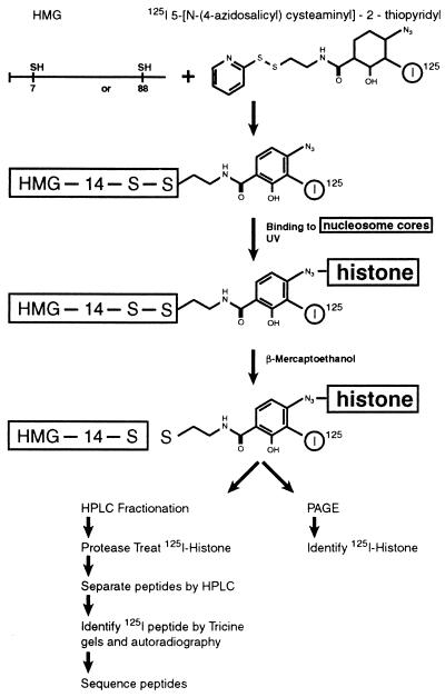
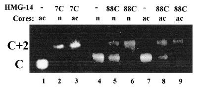
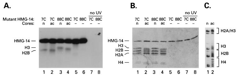
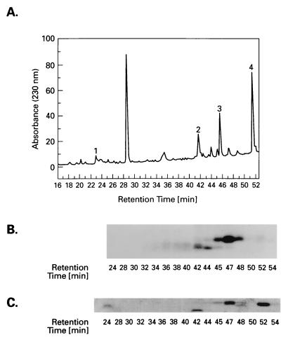
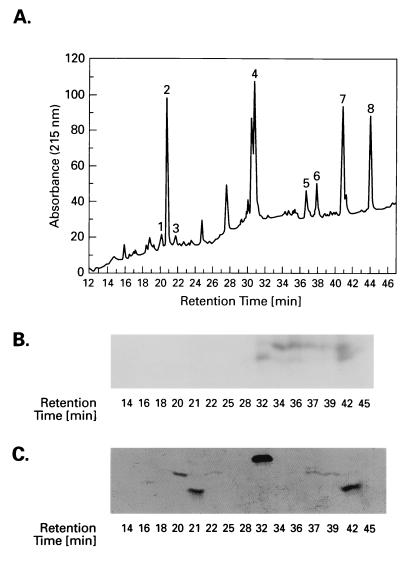
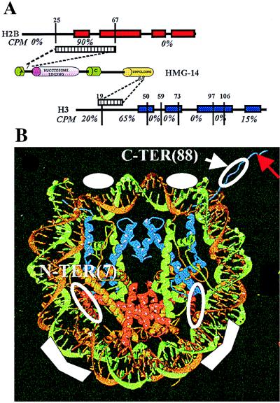
Nonhistone chromosomal protein HMG-14 is a nucleosomal binding protein that unfolds the higher-order chromatin structure and enhances the transcriptional potential of chromatin, but not that of DNA. Both the transcriptional enhancement and the chromatin unfolding activities of HMG-14 are mediated through the C-terminal region of the protein. Here we study the molecular interactions of both this region and the N-terminal region of HMG-14 with nucleosome cores. By protein photocrosslinking we demonstrate that the N-terminal domain of HMG-14 targets a restricted region in histone H2B, whereas the C-terminal chromatin unfolding domain of HMG-14 targets a restricted region in the N terminus of histone H3. The N-terminal regions of the core histones are involved in the folding of oligonucleosomes and are the target of various activities associated with chromatin unfolding and transcriptional activation. We suggest that specific interactions between the C-terminal domain of HMG-14 and the N-terminal tail of histone H3 reduce the compaction of chromatin. These findings provide insights into the molecular mechanism whereby HMG-14/-17 proteins reduce the repressive effect of chromatin, and they also broaden the scope of the molecular interactions involving the N termini of the core histones in nucleosomes.
Figures
Similar articles
-
Interaction of high mobility group-I (Y) nonhistone proteins with nucleosome core particles.J Biol Chem. 1993 Oct 5;268(28):21137-46. J Biol Chem. 1993. PMID: 8407950
-
Alleviation of histone H1-mediated transcriptional repression and chromatin compaction by the acidic activation region in chromosomal protein HMG-14.Mol Cell Biol. 1997 Oct;17(10):5843-55. doi: 10.1128/MCB.17.10.5843. Mol Cell Biol. 1997. PMID: 9315642 Free PMC article.
-
Modular structure of chromosomal proteins HMG-14 and HMG-17: definition of a transcriptional enhancement domain distinct from the nucleosomal binding domain.Mol Cell Biol. 1995 Dec;15(12):6663-9. doi: 10.1128/MCB.15.12.6663. Mol Cell Biol. 1995. PMID: 8524231 Free PMC article.
-
The HMG-14/-17 chromosomal protein family: architectural elements that enhance transcription from chromatin templates.Semin Cell Biol. 1995 Aug;6(4):247-55. doi: 10.1006/scel.1995.0033. Semin Cell Biol. 1995. PMID: 8562917 Review.
-
Intra- and inter-nucleosome interactions of the core histone tail domains in higher-order chromatin structure.Chromosoma. 2014 Mar;123(1-2):3-13. doi: 10.1007/s00412-013-0435-8. Epub 2013 Aug 31. Chromosoma. 2014. PMID: 23996014 Free PMC article. Review.
Cited by
-
Functional interplay between histone H1 and HMG proteins in chromatin.Biochim Biophys Acta. 2016 Mar;1859(3):462-7. doi: 10.1016/j.bbagrm.2015.10.006. Epub 2015 Oct 8. Biochim Biophys Acta. 2016. PMID: 26455954 Free PMC article. Review.
-
Chromosomal proteins HMG-14 and HMG-17 are released from mitotic chromosomes and imported into the nucleus by active transport.J Cell Biol. 1998 Dec 14;143(6):1427-36. doi: 10.1083/jcb.143.6.1427. J Cell Biol. 1998. PMID: 9852141 Free PMC article.
-
Mitotic phosphorylation of chromosomal protein HMGN1 inhibits nuclear import and promotes interaction with 14.3.3 proteins.Mol Cell Biol. 2002 Oct;22(19):6809-19. doi: 10.1128/MCB.22.19.6809-6819.2002. Mol Cell Biol. 2002. PMID: 12215538 Free PMC article.
-
Regulation of chromatin structure and function by HMGN proteins.Biochim Biophys Acta. 2010 Jan-Feb;1799(1-2):62-8. doi: 10.1016/j.bbagrm.2009.11.016. Epub 2009 Nov 27. Biochim Biophys Acta. 2010. PMID: 19948260 Free PMC article. Review.
-
Mammalian transcription-coupled excision repair.Cold Spring Harb Perspect Biol. 2013 Aug 1;5(8):a012625. doi: 10.1101/cshperspect.a012625. Cold Spring Harb Perspect Biol. 2013. PMID: 23906714 Free PMC article. Review.
References
-
- van Holde K E. Chromatin. New York: Springer; 1988.
-
- Wolffe A P. Chromatin Structure and Function. London: Academic; 1995.
-
- Kornberg R D, Lorch Y. Curr Opin Cell Biol. 1995;8:371–375. - PubMed
-
- Adams C C, Workman J L. Cell. 1993;72:305–308. - PubMed
-
- Paranjape S M, Kamakaka R T, Kadonaga J T. Annu Rev Biochem. 1994;63:265–297. - PubMed
MeSH terms
Substances
LinkOut - more resources
Full Text Sources
Miscellaneous
