. 1998 May 12;95(10):5556-61.
doi: 10.1073/pnas.95.10.5556.

Signal-dependent translation of a regulatory protein, Bcl-3, in activated human platelets

Affiliations

Signal-dependent translation of a regulatory protein, Bcl-3, in activated human platelets

A S Weyrich et al. Proc Natl Acad Sci U S A. .

Abstract

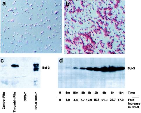
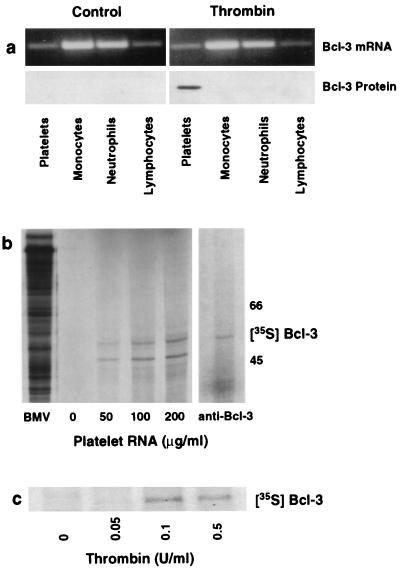
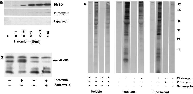
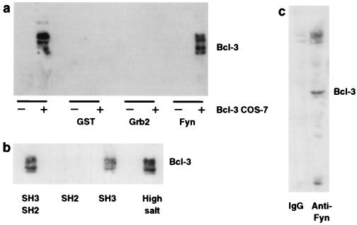
Circulating human platelets lack nuclei, cannot synthesize mRNA, and are considered incapable of regulated protein synthesis. We found that thrombin-activated, but not resting, platelets synthesize Bcl-3, a member of the IkappaB-alpha family of regulatory proteins. The time- and concentration-dependent generation of Bcl-3 in platelets signaled by thrombin was blocked by translational inhibitors, by rapamycin, and by inhibitors of phosphatidylinositol-3-kinase, indicating that it occurs via a specialized translational control pathway that involves phosphorylation of the inhibitory protein 4E-BP1. After its synthesis in activated platelets Bcl-3 binds to the SH3 domain of Fyn (p59(fyn)), a Src-related tyrosine kinase. This, along with its expression in anucleate cells, suggests that Bcl-3 has previously unrecognized functions aside from modulation of transcription. We also demonstrate that platelets synthesize and secrete numerous proteins besides Bcl-3 after they adhere to fibrinogen, which mediates adhesion and outside-in signaling of these cells by engagement of alphaIIb/beta3 integrin. Taken together, these data demonstrate that regulated synthesis of proteins is a signal-dependent activation response of human platelets.


Figures

Figure 1
Figure 1
Activated platelets express Bcl-3. (a and b) Immunocytochemical detection of Bcl-3. Bcl-3 was examined in resting (a) or thrombin-stimulated (b) (0.1 unit/ml for 2 hr) platelets as described in Materials and Methods. Red staining in b indicates the presence of Bcl-3 and is prominent in platelet aggregates. Rabbit IgG, or deletion of the primary antibody, did not result in red staining, indicating that the immunolocalization observed was specific for Bcl-3. (c) Detection of Bcl-3 by Western blot analysis in stimulated platelets and transfected cells. Cellular lysates from control or activated platelets (plts) stimulated with 0.1 unit/ml of thrombin for 2 hr, or from COS-7 cells transfected with vector or Bcl-3 cDNA, were obtained, and Bcl-3 expression (56-kDa protein) was determined by Western blot analysis. In transfected COS-7 cells (lane 4), the slower-migrating bands are phosphorylated forms of Bcl-3 protein that are concentrated in the cytoplasm whereas the faster-migrating band is not phosphorylated and concentrates in the nucleus (not shown). (d) Bcl-3 protein increases over time in platelets activated with thrombin. Platelets were stimulated with thrombin (0.1 unit/ml), and Bcl-3 expression was examined by Western blot analysis over 18 hr. Numbers at the bottom indicate the increase in Bcl-3 protein expression over baseline as measured by densitometry (m, minutes; h, hours). This figure is representative of three experiments.
Figure 2
Figure 2
Platelets synthesize Bcl-3 protein from preexisting mRNA. (a) Thrombin stimulates Bcl-3 protein expression in platelets but not in other blood cells. Platelets and leukocyte subpopulations were isolated, each cell population was exposed to thrombin (0.1 unit/ml) for 2 hr, and total RNA and protein were isolated for reverse transcription–PCR (Upper) and Western blot analysis (Lower). We found the same result in three experiments. (b) In vitro translation of total RNA from platelets results in Bcl-3 protein synthesis. (Left) Increasing concentrations of platelet RNA (μg/ml) were translated in vitro in the presence of [35S]methionine and subsequently examined by autoradiography. mRNA from Brome mosaic virus (BMV) served as a positive control. (Right) The [35S]methionine-labeled protein from platelet RNA translated in vitro was immunoprecipitated with an anti-Bcl-3 antibody as described in Materials and Methods. A second experiment yielded the same result. (c) Radiolabeled incorporation of [35S]methionine into Bcl-3 in platelets activated with thrombin. Platelets were preincubated with [35S]methionine and stimulated with increasing concentrations of thrombin, and lysates were incubated with an anti-Bcl-3 antibody after 4 hr. This figure is representative of three experiments.
Figure 3
Figure 3
Synthesis of Bcl-3 and other proteins is regulated by rapamycin in signaled platelets. (a) Inhibition of translation markedly attenuates Bcl-3 expression in platelets signaled by thrombin. Platelets were pretreated for 2 hr with dimethyl sulfoxide, puromycin, or rapamycin and activated with increasing concentrations of thrombin for 2 hr, and newly synthesized Bcl-3 was identified by Western blot analysis. (b) Rapamycin inhibits 4E-BP1 phosphorylation in activated platelets. Platelets were pretreated with rapamycin, and 4E-BP1, a protein that migrates in SDS/PAGE as multiple species (20–24 kDa), was identified by Western blot analysis 15 min after stimulation with thrombin (0.01 unit/ml). The solid arrow indicates a phosphorylated form of 4E-BP1 as described by Lin and Lawrence (11). a and b are representative of five experiments. (c) Platelets stimulated by adhesion to fibrinogen synthesize proteins. Platelets were pretreated with dimethyl sulfoxide, puromycin, or rapamycin. After pretreatment, they were incubated with [35S]methionine and allowed to adhere to immobilized fibrinogen for 8 hr. Supernatants were removed and the platelets were washed extensively with M199 buffer containing methionine, lysed, and fractionated into soluble and insoluble portions, and the labeled proteins were separated in a 15% SDS-polyacrylamide gel. This experiment is representative of three others.
Figure 4
Figure 4
Bcl-3 binds Fyn in transfected cells and activated platelets. (a) COS-7 cells, transfected with vector or Bcl-3 cDNA, were incubated alone or with agarose-conjugated fusion proteins consisting of GST, GST-Grb2, or GST-Fyn for 2 hr. After extensive washing, protein was eluted from the beads and Bcl-3 was detected by Western blot analysis. (b) Bcl-3 protein expression was detected in cellular lysates from COS-7 cells transfected with Bcl-3 cDNA and subsequently incubated with GST-Fyn fusion proteins that contained either its SH2-SH3 domains, the SH2 domain, or the SH3 domain. Binding of Bcl-3 to the GST-Fyn fusion protein containing both the SH2-SH3 domains was also examined under more stringent conditions (high salt = 400 mM, right lane). (c) Platelets were activated with thrombin (0.1 unit/ml for 2 hr), the cellular lysates were immunoprecipitated with an antibody against Fyn or mouse IgG, and Bcl-3 protein was detected by Western blot analysis as described in Materials and Methods. This figure is representative of four experiments.

References

    1. Elstad M R, McIntyre T M, Prescott S M, Zimmerman G A. Curr Opin Hematol. 1995;2:47–54. - PubMed
    1. Coughlin S R, Vu T-K H, Hung D T, Wheaton V I. J Clin Invest. 1992;89:351–355. - PMC - PubMed
    1. Packham M A. Can J Physiol Pharmacol. 1994;72:278–284. - PubMed
    1. Savage B, Sladivar E, Ruggeri Z M. Cell. 1996;84:289–297. - PubMed
    1. Weksler B B. In: Inflammation: Basic Principles and Clinical Correlates. 2nd Ed. Gallin J I, Goldstein I M, Snyderman R, editors. New York: Raven; 1992. pp. 727–746.

Publication types

MeSH terms

LinkOut - more resources