. 1998 May 12;95(10):5595-600.
doi: 10.1073/pnas.95.10.5595.

Fas-induced proteolytic activation and intracellular redistribution of the stress-signaling kinase MEKK1

Affiliations

Fas-induced proteolytic activation and intracellular redistribution of the stress-signaling kinase MEKK1

J C Deak et al. Proc Natl Acad Sci U S A. .

Abstract

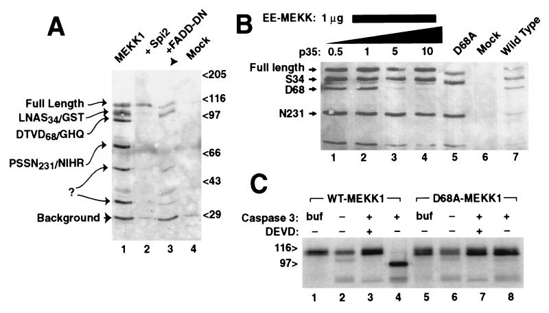
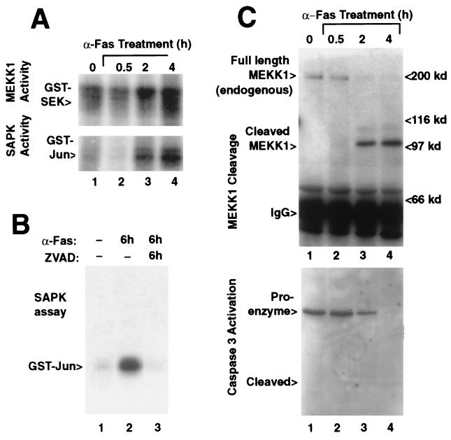
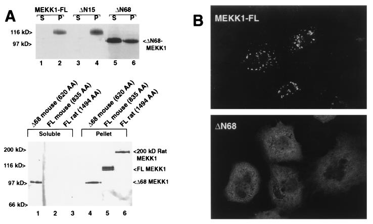
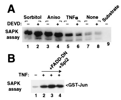
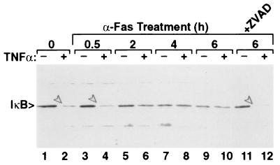
The stress-activated protein kinase (SAPK, alternatively JNK) is activated rapidly by cell stress stimuli such as inflammatory cytokines and oxidative stress, and more slowly by the initiation of the apoptotic cell death response by events such as ligation of the Fas protein. Mitogen-activated protein kinase/Erk kinase kinase-1 (MEKK1) is an activator of SAPK, serving as a SAPK-kinase-kinase through intermediate phosphorylation of the SAPK kinase SEK1. By sequencing proteolytic cleavage products of MEKK1, we found that the proapoptotic protease caspase 3 (CPP32) cleaves MEKK1 after residue D68 both in vivo and in vitro. Cleavage of MEKK1 after D68 is blocked by viral and chemical protease inhibitors. Cleavage of MEKK1 at D68 changes the intracellular distribution of the protein from a Triton-insoluble compartment to a Triton-soluble compartment, reflected in a redistribution from a particulate to a diffuse cytoplasmic staining seen by immunofluorescence. Activation of both SAPK and MEKK1 after Fas ligation is prevented by both viral and chemical caspase 3 inhibitors, which in contrast fail to block activation of SAPK by rapidly acting cell stresses. Stress factor-induced SAPK signaling is not dependent on caspase 3 function. We propose that two mechanisms of stress signaling through MEKK1 exist. One is rapid, independent of proteases, and occurs in the particulate Triton-insoluble compartment. The other is more slowly activated and involves liberation of particulate MEKK1 by proteolytic cleavage and activation by caspase 3.


Figures

Figure 1
Figure 1
MEKK1 is cleaved at amino acid D68 by caspase 3/CPP32. (A) C-terminally GST-tagged MEKK1 expressed in 293 cells demonstrated six plasmid derived bands (lane 1) detected here by glutathione precipitation and anti-GST immunoblotting. Amino terminal sequencing of these fragments confirmed proteolysis at the indicated sites, following amino acids S34, D68, and N231; two other cleavage sites were not determined. Cotransfection of either the vaccinia virus protease inhibitor Spi-2 (lane 2) or dominant negative Δ1–79 FADD (lane 3) prevented cleavage at D68 and smaller bands. (B) Cleavage of MEKK at D68 was blocked by cotransfection of increasing molar ratios of plasmid encoding baculoviral caspase inhibitor p35 (0.5–10 μg), with minimal effects on other cleavage fragments (lanes 1–4). Mutation of codon D68 of MEKK1 to alanine prevented cleavage at codon 68 (lane 5). (C) Cleavage of MEKK1 by caspase 3 in vitro. Full-length murine MEKK1, either wild-type (lanes 1–4) or mutant D68A (lanes 5–8) expressed by using in vitro transcription/translation was treated with buffer alone (lanes 1 and 5) or with extracts of untransformed bacterial cells (lanes 2 and 6), or bacterial extracts expressing active caspase 3 (lanes 3, 4, 7, and 8). Caspase 3 cleaved MEKK1, yielding a 100-kDa band. The caspase inhibitor z-DEVD-fmk blocked the cleavage (lane 3), and the D68A mutant was resistant to cleavage (lane 8).
Figure 2
Figure 2
SAPK and MEKK1 activity correlate with the onset of caspase 3 activation after anti-Fas-induced apoptosis. (A) MEKK1 (Upper) and SAPK (Lower) activity in anti-Fas-treated Jurkat cells gradually increased over a time course of 4 hr (Upper). (B) The activation of SAPK during 6-hr anti-Fas-treatment (lane 2) was blocked by z-VAD-fmk beginning at 15 min before addition of the antibody (lane 3). (C) Cleavage of endogenous MEKK1 in anti-Fas-treated Jurkat cells (Upper) was evidenced by a 100-kDa MEKK1 form detected after 2 hr that was absent in the untreated cells (lane 1). MEKK was detected by immunoprecipitation followed by immunoblotting against MEKK1. Caspase 3 (CPP32) proteolytic activation (Lower) from the same cell lysates correlate with appearance of the proteolysed MEKK1 fragment. Caspase 3 was detected by immunoblotting.
Figure 3
Figure 3
(A) (Upper) MEKK is tethered to a Triton-insoluble compartment by amino acids between 15 and 68. MEKK1 was expressed in CV1 cells by using the T7-vaccinia virus protocol to eliminate proteolytic cleavage. Both full-length (lanes 1 and 2) and ΔN15 MEKK (lanes 3 and 4) were found exclusively in the particulate (P) fraction, and not in the Triton soluble (S) fraction. Mutant ΔN68 that mimics cleavage of MEKK at D68 was primarily found in the soluble fraction. (Lower) Full-length mouse MEKK1 (120 kDa) or Δ68 MEKK1 as in the upper panel were compared with the 200-kDa MEKK1 form isolated from rat. The 200-kDa rat full-length MEKK1 also was found predominantly in the Triton-insoluble compartment. (B) Redistribution of truncated MEKK1 detected by immunofluorescence. Wild-type MEKK1 expressed by using the T7-vaccinia virus system was observed 4 hr after infection in coarse cytoplasmic particles (Upper). Deletion mutant ΔN68 MEKK (Lower) was detected in the cytoplasm and demonstrated a fine reticular pattern. EE-epitope tagged MEKK1 was detected by using anti-EE mAb and confocal imaging.
Figure 4
Figure 4
Conventional stress agents activate SAPK through protease independent pathways. (A) Activation of endogenous SAPK by hypertonic sorbitol (lanes 1 and 2), anisomycin (lanes 3 and 4), or TNFα (lanes 5 and 6) after 30 min was not inhibited by preincubation with the caspase inhibitor z-DEVD-fmk (500 nM, even lanes). (B) Epitope-tagged SAPK expressed in 293 cells was stimulated by TNFα (lane 2) and was not suppressed by coexpression of dominant negative FADD or Spi-2 (lanes 3 and 4). SAPK activity was determined by immunoprecipitation of endogenous SAPK (A) or epitope-tagged p54 SAPK (B) and in vitro kinase reaction by using GST-Jun as substrate.
Figure 5
Figure 5
Fas-stimulated Jurkat cells are insensitive to TNFα-induced IκB degradation. Jurkat cells were stimulated with anti-Fas for the indicated times. Some cultures (even lanes) were treated with 30 ng/ml TNFα for 30 min before harvest. IκB was detected by immunoblotting. Arrows in lanes 2, 4, and 12 denote IκB degraded in response to TNFα. Degradation was hindered by 2 or more hours of anti-Fas treatment. z-VAD inhibition of caspases (lanes 11 and 12) allowed IκB degradation in response to TNFα.
Figure 6
Figure 6
Model of caspase-dependent and independent activation of kinase signaling through MEKK1-SAPK. (Upper) MEKK1 is anchored in a Triton-insoluble compartment within the cell. Cell stressors acting acutely stimulate MEKK1 and SAPK without disrupting this compartment and recognize downstream substrates. Parallel activation of NFκB results in an anti-apoptotic signal. (Lower) Fas-induced activation of caspase 3 or related proteases cleave MEKK1 at codon D68 and release it from the Triton-insoluble compartment. Substrates recognized by the soluble kinase may be distinct from stress-induced substrates and may mediate some aspects of the apoptotic phenotype. TNFα induced activation of NFκB via degradation of IκB is prevented, because of either cleavage of MEKK1 or another uncharacterized mechanism.

References

    1. Kyriakis J M, Banerjee P, Nikolakaki E, Dai T, Rubie E A, Ahmad M F, Avruch J, Woodgett J R. Nature (London) 1994;369:156–160. - PubMed
    1. Kyriakis J M, Avruch J. J Biol Chem. 1996;271:24313–24316. - PubMed
    1. Waskiewicz A J, Cooper J A. Curr Opin Cell Biol. 1995;7:798–805. - PubMed
    1. Lange-Carter C A, Pleiman C M, Gardner A M, Blumer K J, Johnson G L. Science. 1993;260:315–319. - PubMed
    1. Sánchez I, Hughes R T, Mayer B, Yee K, Woodgett J R, Avruch J, Kyriakis J M, Zon L I. Nature (London) 1994;372:795–798. - PubMed

Publication types

MeSH terms