. 1998 May 12;95(10):5611-5.
doi: 10.1073/pnas.95.10.5611.

Development of normal mice from metaphase I oocytes fertilized with primary spermatocytes

Affiliations

Development of normal mice from metaphase I oocytes fertilized with primary spermatocytes

A Ogura et al. Proc Natl Acad Sci U S A. .

Abstract

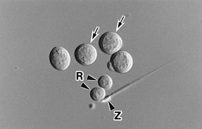
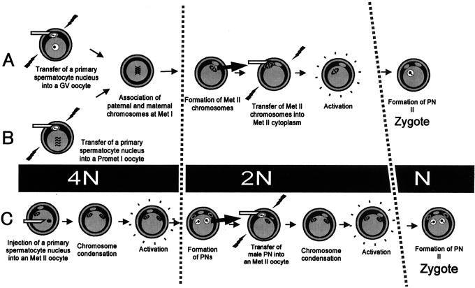
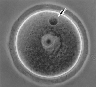
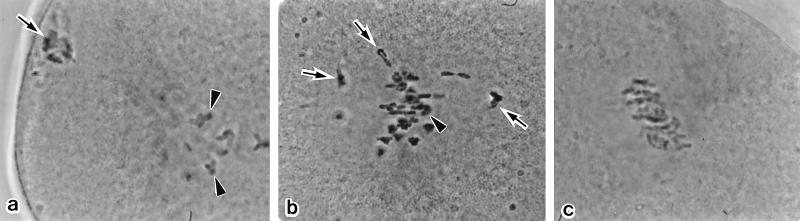
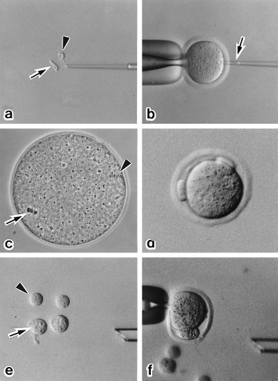
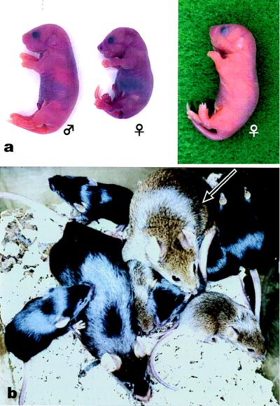
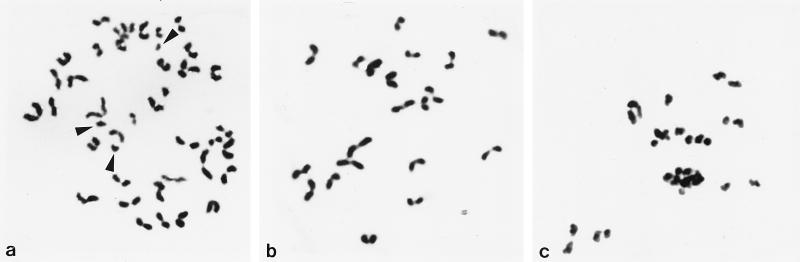
Primary spermatocytes are the male germ cells before meiosis I. To examine whether these 4n diploid cells are genetically competent to fertilize oocytes and support full embryo development, we introduced the nuclei of pachytene/diplotene spermatocytes into oocytes that were arrested in prophase I (germinal vesicle stage), metaphase I, or metaphase II (Met II). Both the paternal and maternal chromosomes then were allowed to undergo meiosis synchronously until Met II. In the first and second groups, the paternal and maternal chromosomes had intermingled to form a large Met II plate, which was then transferred into a fresh enucleated Met II oocyte. In the third group, the paternal Met II chromosomes were obtained by transferring spermatocyte nuclei into Met II oocytes twice. After activation of the Met II oocytes that were produced, those microfertilized at metaphase I showed the best developmental ability in vitro, and three of these embryos developed into full-term offspring after embryo transfer. Two pups (one male and one female) were proven to be fertile. This finding provides direct evidence that the nuclei of male germ cells acquire the ability to fertilize oocytes before the first meiotic division.


Figures

Figure 1
Figure 1
Isolated mouse primary spermatocytes (arrows). Only those with a distinct nuclear membrane were used in this study. R, round spermatid; Z, spermatozoon.
Figure 2
Figure 2
Methods of producing diploid embryos by using primary spermatocytes as male gametes. “N” indicates the number of chromatid sets from primary spermatocytes. The cell-cycle stages of primary spermatocytes and oocytes were synchronized at the GV (Method A), Met I (Method B), or Met II (Method C) stages. Spermatocyte chromosomes (4N) undergo two meiotic divisions in oocytes and participate in the formation of a haploid (N) male pronucleus. PN, pronucleus.
Figure 3
Figure 3
A GV-arrested oocyte fused with a primary spermatocyte (Method A) 4 hr after electrofusion. The spermatocyte nucleus shows its original chromatin configuration (arrow).
Figure 4
Figure 4
The configuration of primary spermatocyte chromosomes in maturing oocytes (Method B). (a) Thirty minutes after electrofusion. The spermatocyte chromosomes have started to condense (arrow). The oocyte chromosomes are at prometaphase I (arrowheads). (b) One hour after electrofusion. The fully condensed spermatocyte chromosomes (arrows) are mingling with the oocyte chromosomes, which already have formed the Met I configuration (arrowheads, slightly out of focus). (c) Two hours after electrofusion. Both the spermatocyte and oocyte chromosomes are aligned on the Met I spindle of the oocyte, which has been arrested by cytochalasin treatment. Thus, the spermatocyte and oocyte chromosomes are successfully synchronized.
Figure 5
Figure 5
Serial nuclear transfer of a spermatocyte nucleus (Method C). (a) The nucleus (arrow) and cytoplasm (arrowhead) of primary spermatocytes are isolated after drawing in and out of the injection pipette. (b) The nucleus of a primary spermatocyte is injected into a Met II oocyte (arrow). (c) The spermatocyte chromosomes form a metaphase plate (arrow) within 2 hr. The arrowhead indicates the Met II chromosomes of the oocyte. (d) After oocyte activation, two polar bodies are extruded. (e) The larger pronucleus (arrow) and polar body (arrowhead) are used for the second nuclear transfer. (f) A karyoplast, pronucleus or polar body, is inserted into the perivitelline space of another Met II oocytes, and a fusion pulse is applied.
Figure 6
Figure 6
Pups born after microfertilization with primary spermatocytes. (a) Just after birth. The smallest pup (female, Center) died soon after birth, although no gross abnormality was found. (b) The male pup grew normally and developed into a fertile adult (arrow).
Figure 7
Figure 7
Chromosome preparation made at Met II. (a) Method A. Some chromatids that separated prematurely show single “I” shapes (arrowheads) whereas normal sister chromatid pairs show characteristic “V” shapes. Oocyte chromosomes are also included in the preparation. (b) Method B. Normal Met II chromosomes (n = 20) derived from a primary spermatocyte. (c) Method C. The spermatocyte chromosomes are very damaged with premature chromatid separation.

References

    1. Ogura A, Matsuda J, Yanagimachi R. Proc Natl Acad Sci USA. 1994;91:7460–7462. - PMC - PubMed
    1. Kimura Y, Yanagimachi R. Development. 1995;121:2397–2405. - PubMed
    1. Sofikitis N V, Miyagawa I, Agapitos E, Pasyianos P, Toda T, Hellstrom W J G, Kawamura H. J Assist Reprod Genet. 1994;11:335–341. - PubMed
    1. Tesarik J, Mendoza C, Testart J. N Engl J Med. 1995;333:525. - PubMed
    1. Kimura Y, Yanagimachi R. Biol Reprod. 1995;53:855–862. - PubMed

Publication types

LinkOut - more resources