. 1998 May 12;95(10):5672-7.
doi: 10.1073/pnas.95.10.5672.

Neuronal defects and delayed wound healing in mice lacking fibroblast growth factor 2

Affiliations

Neuronal defects and delayed wound healing in mice lacking fibroblast growth factor 2

S Ortega et al. Proc Natl Acad Sci U S A. .

Abstract

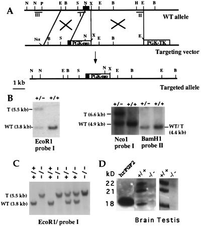
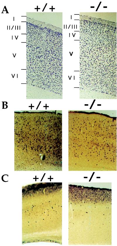
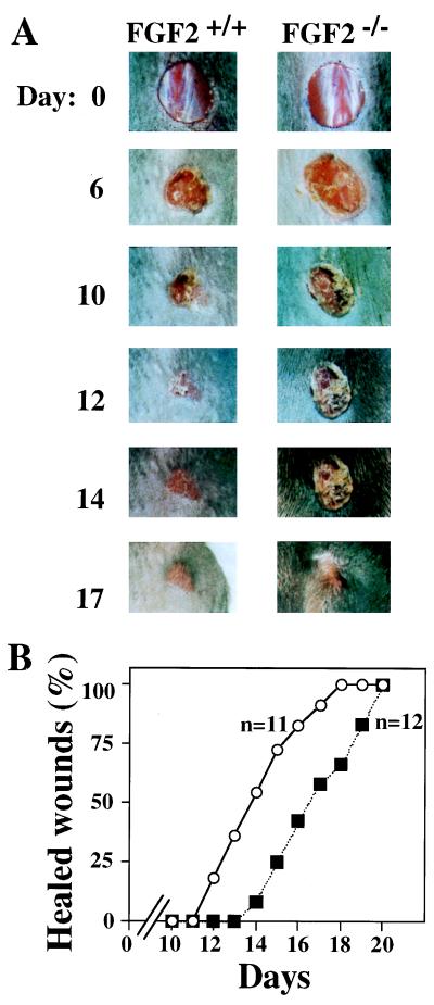
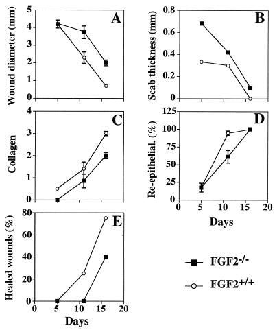
Basic fibroblast growth factor (FGF2) is a wide-spectrum mitogenic, angiogenic, and neurotrophic factor that is expressed at low levels in many tissues and cell types and reaches high concentrations in brain and pituitary. FGF2 has been implicated in a multitude of physiological and pathological processes, including limb development, angiogenesis, wound healing, and tumor growth, but its physiological role is still unclear. To determine the function of FGF2 in vivo, we have generated FGF2 knockout mice, lacking all three FGF2 isoforms, by homologous recombination in embryonic stem cells. FGF2(-/-) mice are viable, fertile and phenotypically indistinguishable from FGF2(+/+) littermates by gross examination. However, abnormalities in the cytoarchitecture of the neocortex, most pronounced in the frontal motor-sensory area, can be detected by histological and immunohistochemical methods. A significant reduction in neuronal density is observed in most layers of the motor cortex in the FGF2(-/-) mice, with layer V being the most affected. Cell density is normal in other regions of the brain such as the striatum and the hippocampus. In addition, the healing of excisional skin wounds is delayed in mice lacking FGF2. These results indicate that FGF2, although not essential for embryonic development, plays a specific role in cortical neurogenesis and skin wound healing in mice, which, in spite of the apparent redundancy of FGF signaling, cannot be carried out by other FGF family members.


Figures

Figure 1
Figure 1
FGF2 gene targeting. (A) Partial map of the FGF2 gene (Top), gene-targeting vector (Middle), and FGF2-targeted allele (Bottom). The first exon of the FGF2 gene is shown as a black box. The neo- and hsvTK-expressing cassettes, containing the PGK-1 promoter and polyadenylation sequences, are shown as white boxes. The position of the relevant restriction sites for the enzymes BamHI (B), EcoRI (E), XbaI (X), NcoI (N), SalI (S), HindIII (H), and PstI (P) is marked. The NotI site was used to linearize the targeting vector. DNA fragments I, II, and III were used as probes in Southern blot hybridization. (B) Genomic DNA from ES cells was digested with EcoRI, NcoI, and BamHI and hybridized to the probes indicated. T, targeted allele. (C) Southern blot of tail DNA from the offspring of heterozygous FGF2+/− mutant parents. DNA was digested with EcoRI and probed with probe I. (D) Absence of FGF2 in protein extracts from brain and testis of FGF2 −/− mice. Western blots of protein extracts concentrated on heparin-Sepharose beads. Blots were probed with a monoclonal anti-FGF-2 antibody (Transduction Laboratories). The same result was obtained with polyclonal antibodies for FGF2. The position of the three isoforms of FGF2 is indicated; 25 ng of human recombinant FGF2 was loaded as a control.
Figure 2
Figure 2
Defects in the motor area of the neo-cortex of FGF2−/− mice. Coronal sections of the FGF2−/− mutant cortex compared with those of wild-type FGF2+/+ cortex after cresyl violet (NISSL) staining (A), parvalbumin (B), and calbindin (C) immunohistochemistry. Reduced cell density is evident in layers V and VI in AC.
Figure 3
Figure 3
Skin wound healing of FGF2−/− mice. (A) Example of the healing progress in an FGF2−/− mouse and an FGF+/+ mouse. Wounds were photographed at the time indicated. Day 0 picture was taken immediately after wounding. All wounds were photographed at the same distance. (B) The fraction of completely healed wounds (determined as described in Materials and Methods) is plotted versus the time after wounding. n, number of mice of each genotype analyzed. Wounds were performed in a genotype-blind fashion. Mice were caged separately throughout the experiment. ○, FGF2+/+; ▪, FGF2−/−.
Figure 4
Figure 4
Histologic analysis of wound healing in FGF2−/− and FGF2+/+ mice. Wound diameter (A), scab thickness (B), collagen deposition (C), percentage of reepithelialization (D), and percentage of completely healed wounds (E) are plotted versus time after wounding. The number of animals was five FGF2−/− and five FGF2+/+ at 5 days and 16 days after wounding, respectively, and seven FGF2−/− and seven FGF2+/+ at day 11. Each point represents the mean ± SEM, except in E. Wounds that were completely healed were not considered in the calculation of the mean. The histologic parameters were determined as described in Materials and Methods.

References

    1. Baird A, Bohlen P. In: Peptide Growth Factors and Their Receptors. Sporn M B, Roberts A B, editors. New York: Springer; 1991. pp. 369–418.
    1. Basilico C, Moscatelli D. Adv Cancer Res. 1992;59:115–165. - PubMed
    1. Bikfalvi A, Klein S, Pintucci G, Rifkin D B. Endocr Rev. 1997;18:26–45. - PubMed
    1. Goldfarb M. Cytokine Growth Factor Rev. 1996;7:311–325. - PubMed
    1. Mansukhani A, Dell’Era P, Moscatelli D, Kornbluh S, Hanafusa H, Basilico C. Proc Natl Acad Sci USA. 1992;89:3305–3309. - PMC - PubMed

Publication types

Substances