. 1998 May 12;95(10):5694-9.
doi: 10.1073/pnas.95.10.5694.

Alternative splicing of rearranged T cell receptor delta sequences to the constant region of the alpha locus

Affiliations

Alternative splicing of rearranged T cell receptor delta sequences to the constant region of the alpha locus

F Livák et al. Proc Natl Acad Sci U S A. .

Abstract

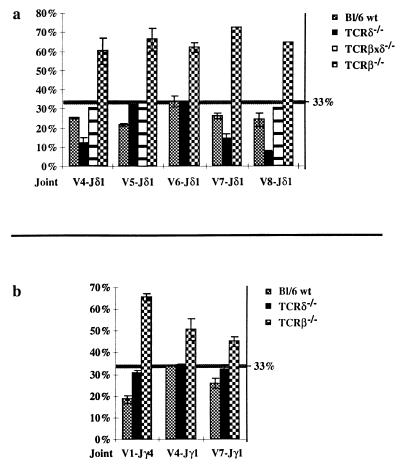
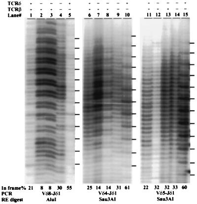
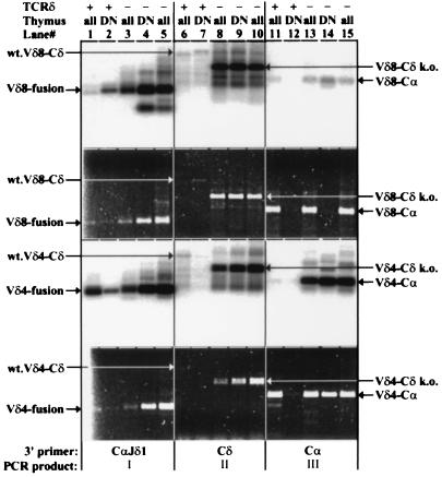
The T cell receptor (TCR) alpha/delta locus is composed of a common, shared set of variable (V) and distinct diversity (D), joining (J), and constant (C) genes. It has been recognized for several years that transcripts of the rearranged VDJdelta or VJalpha genes are spliced to the Cdelta or Calpha genes, respectively, encoding distinct TCR delta and alpha proteins. Herein, we describe the discovery of a splicing variation that allows the assembled VDJdelta genes to be fused with the Calpha gene. This variation is prominent in TCRdelta gene-deficient mice but is also detectable in wild-type mice. Furthermore, we show that several in-frame VDJdelta rearrangements in TCRdelta gene-deficient mice are strikingly underrepresented, suggesting that the alternative transcripts, with protein coding capacity, influence the development of alphabeta thymocytes. In-frame TCRgamma gene rearrangements do not appear underrepresented, indicating that the effect is not mediated by the gamma chain. Instead, indirect evidence supports the hypothesis that the delta/alpha chimeric protein acts in conjunction with the TCRbeta chain. These results have implications for the transcriptional control of the TCRalpha/delta locus and provide a novel insight into the distinct functional capacities of the TCR alpha and delta proteins during thymocyte development.


Figures

Figure 1
Figure 1
(a) Schematic map of the TCRα/δ locus (not to scale). Boxes represent the various coding sequences. For the C genes, boxes are used to indicate each of the four exons. Arrows within the V gene boxes indicate transcriptional orientation. The top and bottom lines depict the wild-type and TCRδ-deficient loci, respectively. (b) An example of a V gene (Vδ4) rearrangement that can generate VDJδ1-Cα transcripts on the TCRδ-deficient locus. Lines indicate the two potential splicing events that can give rise to the two distinct mRNA species shown below the locus. Solid lines indicate the three RT–PCR products (marked as I, II, and III) that are detected in Fig. 4. (c) An example of a V gene (Vδ5) rearrangement that cannot generate VDJδ1-Cα transcripts on the TCRδ-deficient locus. Note the reverse transcriptional orientation of the assembled Vδ5DJδ1-Cδ region generated by the chromosomal inversion. Lines indicate the only potential splicing event that can give rise to the Vδ5DJδ1-Cδ mRNA shown below the locus and the solid line indicates the only RT–PCR product (marked as II) that can be detected (data not shown).
Figure 2
Figure 2
Graphical representation of quantitative analyses of PCR RFLP reactions on various Vδ-DJδ1 (a) and Vγ-Jγ (b) rearrangements in total thymus samples from C57BL/6 wild-type (Bl/6 wt), TCRδ-deficient (δ−/−), TCRβxδ-deficient (βxδ−/−, only for Vδ-DJδ1), and TCRβ-deficient (β−/−) mice. The percent of in-frame rearrangements is shown on the vertical axes. The position of the 33% random level is marked with a horizontal line. The error bars indicate the standard deviation from the mean for each joint where at least two independent samples were analyzed. Some individual samples were measured several times on different gels.
Figure 3
Figure 3
PCR RFLP analysis of Vδ4, Vδ5, and Vδ8 to DJδ1 rearrangements. DNA samples from total thymi with the indicated genotype (+/+ or −/− for the TCRδ and/or TCRβ loci) were PCR-amplified and digested with the indicated restriction enzymes. Dashes on the right mark the position of the in-frame joints as determined from TCRβ-deficient mice which have only γδ TCR selected cells (14). The percentage of in-frame rearrangements is shown below each lane. Two individual TCRδ samples are shown.
Figure 4
Figure 4
RT–PCR analysis of VDJδ transcripts from wild-type (TCRδ+) and TCRδ-deficient (TCRδ−) total (all) or sorted HSAhi, CD25+, CD4/CD8 double negative (DN) thymocytes. The first and third panels from the top show hybridization to a Jδ1 gene-specific oligonucleotide probe. The second and fourth panels show the corresponding gels stained with ethidium bromide and visualized with UV illumination. Two independent TCRδ total thymus samples are shown. The top two panels show results obtained with a Vδ8-specific forward primer, and the bottom two show results with a Vδ4-specific forward primer. Lanes: 1–5, PCRs performed with the 3′CαJδ1 hybrid reverse primer; 6–10, PCRs performed with a Cδ reverse primer; 11–15, PCRs performed with a Cα reverse primer. The predominant PCR product (I, II, or III, as defined in Fig. 1) for each group of five lanes is indicated at the bottom. The discrepancy observed between the hybridization and ethidium staining signals for Vδ8 RT–PCR reactions in lanes 1 vs. 2, lanes 4 vs. 5, and lanes 13 and 15 vs. 14 is probably due to mispriming (lanes 1–5) or priming (lanes 13–15) of RT–PCR from VJα-Cα templates abundant in total thymus but absent from DN thymus templates. Note the difference in the size of the Cδ-specific RT–PCR products between wild-type and TCRδ-deficient mice due to the targeted deletion of the Cδ sequences. The identity of these bands was assigned based on their expected sizes and for the Vδ4-Cδ products from TCRδ-deficient samples was also confirmed by DNA sequencing.

References

    1. Schatz D G, Oettinger M A, Schlissel M S. Annu Rev Immunol. 1992;10:359–383. - PubMed
    1. Chien Y, Iwashima M, Kaplan K B, Elliot J F, Davis M M. Nature (London) 1987;327:677–682. - PubMed
    1. Wang K, Klotz J L, Kiser G, Bristol G, Hays E, Lai E, Gese E, Kronenberg M, Hood L. Genomics. 1994;20:419–428. - PubMed
    1. Kronenberg M, Siu G, Hood L, Shastri N. Annu Rev Immunol. 1986;4:529–591. - PubMed
    1. Koop B F, Wilson R K, Wang K, Vernooij B, Zallwer D, Kuo C, Seto D, Toda M, Hood L. Genomics. 1992;13:1209–1230. - PubMed

Publication types

MeSH terms

Substances

LinkOut - more resources