. 1998 May 12;95(10):5718-23.
doi: 10.1073/pnas.95.10.5718.

Activation of NF-kappaB via a Src-dependent Ras-MAPK-pp90rsk pathway is required for Pseudomonas aeruginosa-induced mucin overproduction in epithelial cells

Affiliations

Activation of NF-kappaB via a Src-dependent Ras-MAPK-pp90rsk pathway is required for Pseudomonas aeruginosa-induced mucin overproduction in epithelial cells

J D Li et al. Proc Natl Acad Sci U S A. .

Abstract

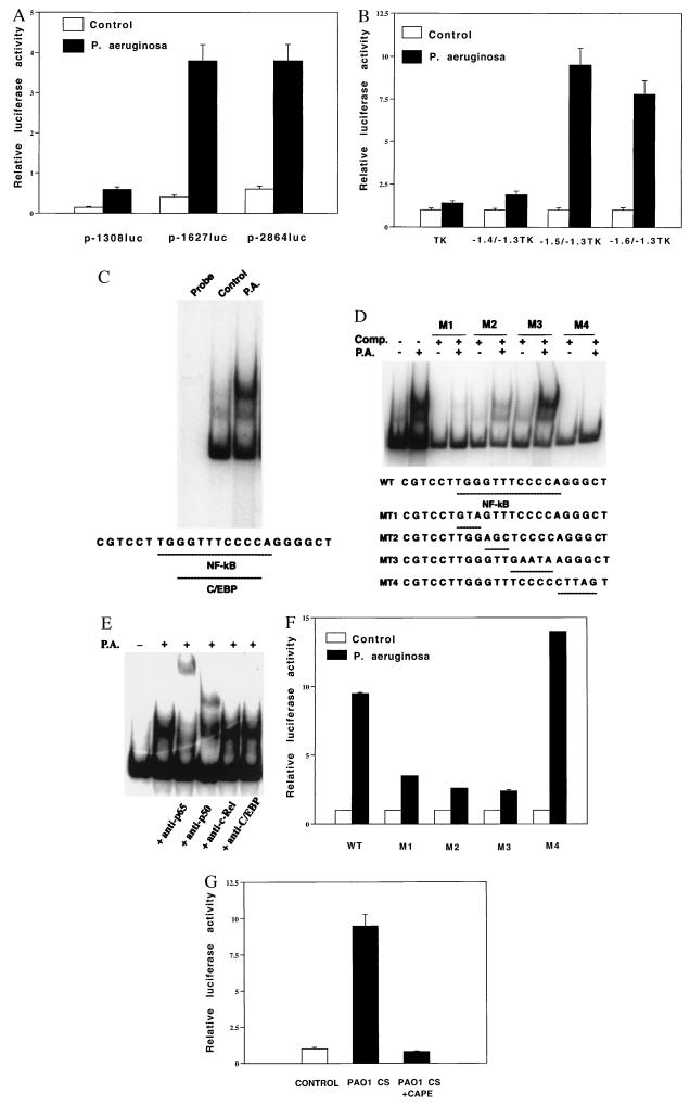
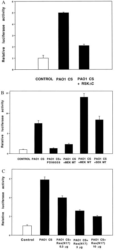
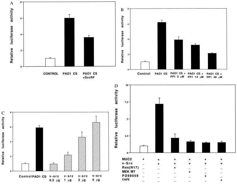
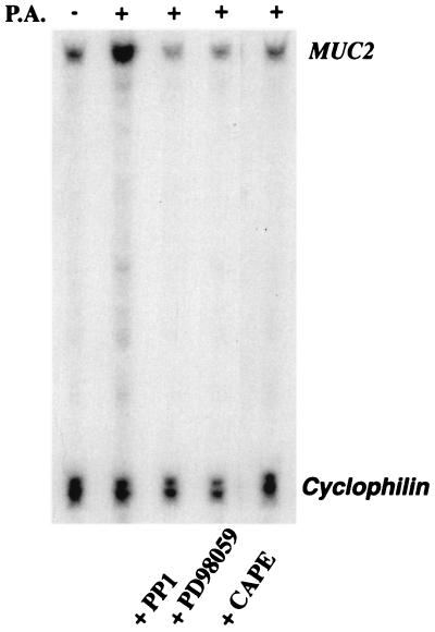
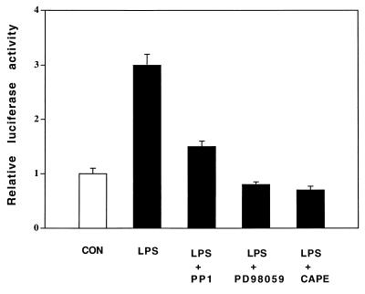
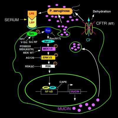
Cystic fibrosis (CF) is an autosomal recessive disorder, the most common lethal genetic disease in Caucasians. Respiratory disease is the major cause of morbidity and mortality. Indeed, 95% of CF patients die of respiratory failure. Pseudomonas aeruginosa, an opportunistic pathogen, chronically infects the lungs of over 85% of CF patients. It is ineradicable by antibiotics and responsible for airway mucus overproduction that contributes to airway obstruction and death. The molecular mechanisms underlying this pathology are unknown. Here we show that P. aeruginosa activates a c-Src-Ras-MEK1/2-MAPK-pp90rsk signaling pathway that leads to activation of nuclear factor NF-kappaB (p65/p50). Activated NF-kappaB binds to a kappaB site in the 5'-flanking region of the MUC2 gene and activates MUC2 mucin transcription. These studies bring new insight into bacterial-epithelial interactions and more specifically into the molecular pathogenesis of cystic fibrosis. Understanding these signaling and gene regulatory mechanisms opens up new therapeutic targets for cystic fibrosis.


Figures

Figure 1
Figure 1
Characterization of NF-κB site and NF-κB in mucin MUC2 induction by P. aeruginosa. (A) Human MUC2 regulatory regions (base pairs −2864 to +14, −1627 to +14, and −1308 to +14) were subcloned upstream of a luciferase reporter gene in pGL2 basic vector and transfected into HM3 cells. (B) Human MUC2 regulatory regions (base pairs −1628 to −1307, −1528 to −1307, and −1430 to −1307) were subcloned upstream of a TK-32 promoter luciferase vector (named as −1.6/−1.3TK, −1.5/−1.3TK, and −1.4/−1.3TK, respectively) and transfected into HM3 cells. In both A and B, transfected cells were treated with either P. aeruginosa culture supernatants or vehicle for 6 hr prior to cell lysis. Luciferase activity was then assessed in P. aeruginosa-treated and nontreated cells. Nuclear proteins were incubated with a probe consisting of the 32P-labeled double-stranded oligonucleotides corresponding to the human MUC2 regulatory region base pairs −1458/−1430 in the absence (C) or presence of various mutant unradiolabeled oligonucleotides (M1 to M4) as indicated (D), or preincubated with antibodies to NF-κB p65, NF-κB p50, c-Rel, or C/EBP (E), and were subjected to EMSA. Probe incubation was with nuclear extracts (10 μg) from HM3 cells either treated or untreated with P. aeruginosa culture supernatant as indicated at the top. (F) Human MUC2 regulatory regions (base pairs −1528 to −1430) containing the wild type (WT) or various mutated sites within the region −1458/−1430 as indicated above were subcloned upstream of the TK-32 promoter in a luciferase vector (named as M1 to M4)) and transfected into HM3 cells. Transfected cells were treated with either P. aeruginosa culture supernatants or vehicle for 6 hr prior to cell lysis. Luciferase activity was then assessed in P. aeruginosa-treated and untreated cells. (G) Effect of NF-κB inhibitor CAPE. A human MUC2 construct p-2864luc was transfected into HM3 cells. Transfected cells were pretreated with or without CAPE (15 μg/ml) for 1 hr before P. aeruginosa treatment as indicated. In all the experiments shown above, transfections were carried out in triplicate. Values are the means ± SD; n = 3. Similar results were observed in another MUC2-expressing epithelial cell line, NCIH292.
Figure 2
Figure 2
Involvement of Ras, MEK1/2, and pp90rsk in MUC2 mucin induction by P. aeruginosa. (A) Effect of coexpressing a dominant-negative mutant form of pp90rsk (pp90rskΔC) on P. aeruginosa-induced MUC2 up-regulation. p-2864luc (15 μg) was cotransfected with a dominant-negative mutant form of pp90rsk into HM3 cells. Forty hours later, HM3 cells were exposed to PAO1 culture supernatant (CS) 6 hr before harvesting. Luciferase activity was measured as described in the text. (B) Effect of PD98059 coexpressing a dominant-negative mutant form and wild-type form of MEK1/2 and a dominant-negative mutant form of SEK [JNKK9(K116R)] on P. aeruginosa-induced MUC2 up-regulation. HM3 cells were cotransfected with p-2864luc (15 μg) and a dominant-negative mutant form (3 μg) or a wild-type form (3 μg) of MEK1/2. Forty hours after transfection, HM3 cells transfected with only p-2864luc were pretreated with PD98059 (37 μM) (Calbiochem) for 30 min. All the cells were then exposed to PAO1 culture supernatant (CS) 6 hr before harvesting. (C) Effect of coexpressing a dominant-negative mutant form of Ras (RasN17) on P. aeruginosa-induced MUC2 up-regulation. HM3 cells were cotransfected with p-2864luc (15 μg) and a dominant-negative mutant form of Ras. The cells were then treated with PAO1 culture supernatant and lysed for luciferase assay. Similar results were observed in another MUC2-expressing epithelial cell line, NCIH292. In the experiments described above, all transfections were carried out in triplicate. Values are the means ± SD; n = 3.
Figure 3
Figure 3
Involvement of Src in MUC2 mucin induction by P. aeruginosa. (A and B) Inhibition of P. aeruginosa-induced MUC2 up-regulation by coexpressing a dominant-negative mutant form of c-Src (A) and Src inhibitor PP1 (B). p-2864luc was transfected into HM3 cells with (A) or without (B) a dominant-negative mutant form of c-Src. Forty hours later, HM3 cells were pretreated with (B) or without PP1 (A) (5, 14, and 28 μM) (Calbiochem) for 30 min and then exposed to PAO1 culture supernatant (CS) for 6 hr before harvesting. Luciferase activity was measured as described in the text. (C) Effect of coexpressing p-2864luc and v-Src. p-2864luc (15 μg) was cotransfected with v-Src (0.3, 1, 3, and 5 μg) into HM3 cells. Forty-eight hours after being transfected, HM3 cells were lysed for assessing luciferase activity. (D) Effect of PD98059, CAPE, and coexpressing a dominant-negative mutant form of Ras and MEK1/2 on v-Src-induced MUC2 up-regulation. p-2864luc (15 μg) was cotransfected with v-Src (3 μg) and a dominant-negative mutant form of either Ras or MEK1/2 into HM3 cells. Forty hours after being transfected, the cells transfected with only p-2864luc were pretreated with either PD98059 (37 μM, 30 min) or CAPE (15 μg/ml, 1 hr). All the cells were then exposed to P. aeruginosa for 6 hr. Forty hours after being transfected, HM3 cells were either exposed or not exposed to PAO1 culture supernatant (CS) 6 hr before harvesting for assessing luciferase activity. Similar results were observed in another MUC2-expressing epithelial cell line, NCIH292. In all the experiments described above, all transfections were carried out in triplicate. Values are the means ± SD; n = 3.
Figure 4
Figure 4
Effects of inhibitors of Src, MEK1/2, and NF-κB on up-regulation of MUC2 mRNA induced by P. aeruginosa. HM3 cells were pretreated with PP1 (14 μM, 30 min), PD98059 (37 μM, 30 min), or CAPE (15 μg/ml, 60 min) and then exposed to PAO1 culture supernatant (CS) for 6 hr before harvesting for RNA extraction. RPA was then performed with the MUC2 gene-specific probe HAM1. A cyclophilin probe was included as a control to assess amount of RNA used in each hybridization reaction. Similar results were observed in another MUC2-expressing epithelial cell line, NCIH292.
Figure 5
Figure 5
Effects of inhibitors of Src, MEK1/2, and NF-κB on up-regulation of MUC2 transcription induced by P. aeruginosa LPS. HM3 cells were transfected with a MUC2 base pair −1,600/−1,300 TK promoter construct. Forty hours later, cells were pretreated with PP1 (14 μM, 30 min), PD98059 (37 μM, 30 min), or CAPE (15 μg/ml, 60 min) and then exposed to P. aeruginosa LPS (15 μg/ml) for 6 hr before harvesting for assessing luciferase activity. Similar results were observed in another MUC2-expressing epithelial cell line, NCIH292.
Figure 6
Figure 6
Schematic diagram showing steps in the signaling pathway by which P. aeruginosa up-regulates human MUC2 mucin gene transcription. As indicated, P. aeruginosa releases LPS, thereby activating a c-Src-Ras-Raf-1-MEK1/2-MAPK (ERK1/2)-pp90rsk pathway, which in turn leads to the activation of NF-κB and triggers MUC2 mucin transcription. LBP, LPS-binding protein. The overproduced mucin, in concert with abnormal airway lining fluid secondary to CFTR mutation, leads to airway mucus obstruction and lung failure.

References

    1. Collins F S, Wilson J M. Nature (London) 1992;358:708–709. - PubMed
    1. Pier G B, Grout M, Zaidi T S, Oslen J C, Johnson L G, Yankasas J R, Goldberg J B. Science. 1996;271:64–67. - PMC - PubMed
    1. Smith J J, Travis S M, Greenberg E P, Welsh M J. Cell. 1996;85:229–236. - PubMed
    1. Saiman L, Prince A. J Clin Invest. 1993;92:1875–1880. - PMC - PubMed
    1. Li J D, Dohrman A F, Gallup M G, Miyata S, Gum J R, Kim Y S, Nadel J A, Price A, Basbaum C B. Proc Natl Acad Sci USA. 1997;94:967–972. - PMC - PubMed

Publication types

MeSH terms