. 1998 May 12;95(10):5746-51.
doi: 10.1073/pnas.95.10.5746.

Host cell dependence of viral morphology

Affiliations

Host cell dependence of viral morphology

P C Roberts et al. Proc Natl Acad Sci U S A. .

Abstract

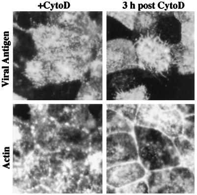
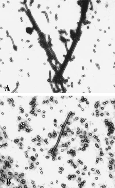
The morphology of influenza virions was found to depend on cellular determinants. Influenza viral filaments up to 30 microm in length were observed to form at high levels on surfaces of various polarized epithelial cell types infected with the A/Udorn/72 virus. In contrast, virions produced by nonpolarized cell types infected with this virus were almost exclusively of spherical morphology. Disruption of the actin microfilament array by cytochalasin D treatment of polarized MDCK cells had a profound effect on viral morphology. Although virus titers and release of spherical particles were not reduced in the presence of cytochalasin D, we observed a 15-fold reduction in the release of filamentous particles. In contrast, the ratio of filaments to spheres produced by infected MDCK cells was not altered by the microtubule-disrupting agent nocodazole. These observations indicate that the polarized cell phenotype and the integrity of the actin microfilament network are important cellular determinants of the morphology of a filamentous influenza virus.


Figures

Figure 1
Figure 1
Visualization of filamentous particle formation in MDCK cells. (a and b) Immunofluorescence (a) and phase-contrast microscopy (b) at 9 h.p.i., revealing the presence of numerous influenza virus filaments emanating from the cell surface. The immunofluorescence shows double staining with HA-specific antibody (red) and antibody to vimentin (green). The latter did not colocalize to viral filaments. (c) Low-magnification electron micrograph showing the presence of many long viral filaments and filament bundles projecting from the surface of A/Udorn-infected MDCK cells. (d and e) Electron micrographs of negatively stained filamentous particles released into the medium. The viral filaments depicted in d are 5–7 μm in length. The characteristic viral spike-like surface projections are readily visible at higher magnification (e). (a and b = ×1,000; c = ×15,000; d = ×10,000; e = ×85,000.)
Figure 2
Figure 2
(a and b) Immunofluorescence micrographs of cell surfaces of Vero C1008 and Vero 76 cells stained with anti-Udorn antisera and FITC-conjugated rabbit anti-guinea pig Ig. Note the intense cell surface staining of Vero 76 cells but lack of filamentous particles (a), whereas filamentous particles (arrows) are budding from infected Vero C1008 cells (b). (c and d) Electron micrographs of negatively stained virus released from BHK-21 cells or HEC-1 cells. Filamentous particles were frequently observed in the virus released from HEC-1 cells (d) but spherical or slightly elongated particles were produced by BHK-21 cells (c). (a and b = ×500; c and d = ×3,800.)
Figure 3
Figure 3
Sucrose gradient separation of spherical and filamentous virus particles. Spherical particles derived from A/Udorn-infected MDCK cells sediment in fractions 11–13, whereas the faster-sedimenting filamentous particles are found in fractions 8–10 (7). The laboratory-adapted strain A/WSN (WSN), which produces strictly spherical particles, sediments in fractions 11–13. Virus derived from infected BHK-21 or from MDCK cells treated with cytochalasin D (10 μg/ml) also predominantly sediments in fractions 11–13.
Figure 4
Figure 4
Inhibition of filament formation and recovery from cytochalasin D treatment. Cells were incubated in the presence of cytochalasin D (CytoD) for 6 hr, the drug was removed, and the infection was allowed to continue at 37°C. At 0 or 3 hr after removal of the inhibitor, cells were doubly stained with guinea pig anti-Udorn antisera and rabbit anti-guinea pig-TRITC (viral antigen) and with FITC-phalloidin (actin). In cytochalasin D-treated cells at 6 h.p.i., intense cell surface staining of viral antigen was observed, but filament formation was not observed. Phalloidin staining confirmed that actin microfilaments were disrupted into aggregates. At 3 h after drug removal, short viral filaments were evident, concomitant with reformation of the actin microfilament array. (×550.)
Figure 5
Figure 5
Effects of cytochalasin D on virus morphology. Negatively stained preparations of A/Udorn virus produced in untreated control MDCK cells (A) and cytochalasin D-treated MDCK cells (B) were examined by electron microscopy. Note the reduction of filamentous particles and abundance of spherical forms present in the cytochalasin D-treated sample. (×8,900.)

References

    1. Ada G L, Perry B T, Abbot A. J Gen Microbiol. 1958;19:23–39. - PubMed
    1. Smirnov Y A, Kuznetsova M A, Kaverin N V. Arch Virol. 1991;118:279–284. - PubMed
    1. Choppin P W. Virology. 1963;21:342–352. - PubMed
    1. Kilbourne E D, Murphy J S. J Exp Med. 1960;111:387–405. - PMC - PubMed
    1. Kilbourne E D. Prog Med Virol. 1963;5:79–126.

Publication types

LinkOut - more resources