. 1998 May 12;95(10):5757-61.
doi: 10.1073/pnas.95.10.5757.

Isolation of a hepadnavirus from the woolly monkey, a New World primate

Affiliations

Isolation of a hepadnavirus from the woolly monkey, a New World primate

R E Lanford et al. Proc Natl Acad Sci U S A. .

Abstract

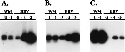
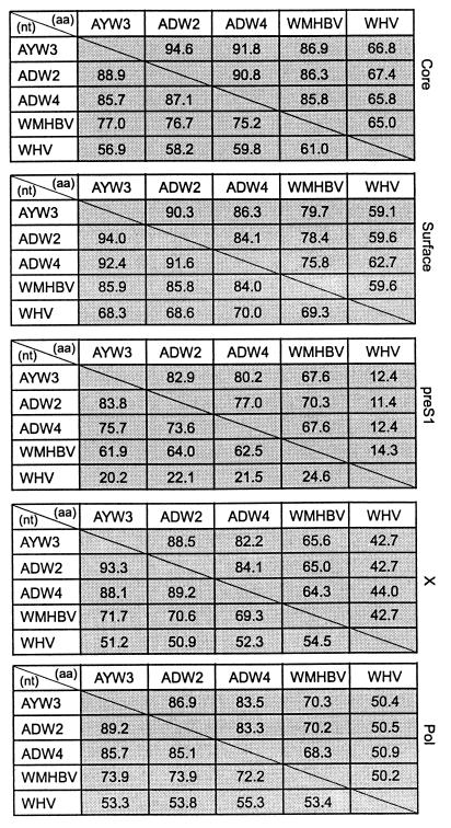
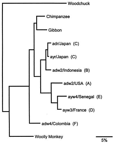
Hepatitis B virus (HBV) infections are a major worldwide health problem with chronic infections leading to cirrhosis and liver cancer. Viruses related to human HBV have been isolated from birds and rodents, but despite efforts to find hepadnaviruses that infect species intermediate in evolution between rodents and humans, none have been described. We recently isolated a hepadnavirus from a woolly monkey (Lagothrix lagotricha) that was suffering from fulminant hepatitis. Phylogenetic analysis of the nucleotide sequences of the core and surface genes indicated that the virus was distinct from the human HBV family, and because it is basal (ancestral) to the human monophyletic group, it probably represents a progenitor of the human viruses. This virus was designated woolly monkey hepatitis B virus (WMHBV). Analysis of woolly monkey colonies at five zoos indicated that WMHBV infections occurred in most of the animals at the Louisville zoo but not at four other zoos in the United States. The host range of WMHBV was examined by inoculation of one chimpanzee and two black-handed spider monkeys (Ateles geoffroyi), the closest nonendangered relative of the woolly monkey. The data suggest that spider monkeys are susceptible to infection with WMHBV and that minimal replication was observed in a chimpanzee. Thus, we have isolated a hepadnavirus with a host intermediate between humans and rodents and establishes a new animal model for evaluation of antiviral therapies for treating HBV chronic infections.


Figures

Figure 1
Figure 1
Southern blot hybridization of PCR products amplified from woolly monkey (lanes WM) serum or HBV containing serum. DNA was extracted from 1:10 dilutions of woolly monkey or HBV containing serum. U, undiluted; −1 to −5 = 10−1 to 10−5 dilutions. PCR was conducted with primers to the core gene. Hybridization of identical membranes was performed with an HBV DNA probe under high (A) or low (B) stringency conditions or with the isolated DNA fragment from amplification of the woolly monkey serum (C) under low-stringency conditions.
Figure 2
Figure 2
Family tree of woolly monkeys from the Louisville Zoo. Shaded circles indicate animals from Scotland. The initials of two sires Herman (H) and Willie (W) are indicated to clarify parentage. ?, sire is unknown; open squares, male; open circles, female; *, index case; + or −, positive and negative for WMHBV markers. Serum samples for some animals were not available for analysis.
Figure 3
Figure 3
Genome organization of WMHBV. The WMHBV genome has the same genetic organization as human HBV. The outer bars show the various HBV ORFs. The inner circle depicts the viral DNA present in the virion that is partially double stranded with the polymerase covalently bound to the 5′ end of the minus strand of DNA and a viral RNA primer at the 5′ end of the plus strand of DNA. The small bars represent the DR1 and DR2 sequences involved in genomic replication.
Figure 4
Figure 4
Comparison of amino acid and nucleotide similarity between hepadnaviruses. The sequences of WMHBV, three human HBV isolates (ayw3/France, genotype D; adw2/Indonesia, genotype B; and adw4/Colombia, genotype F), and woodchuck HBV are compared for percent similarity by using the megalign program of lasergene. Comparisons of the ORFs of the core, surface, pre-S1, X, and polymerase (pol) are shown. Numbers above and below the diagonal line are percent amino acid and nucleotide similarity, respectively.
Figure 5
Figure 5
Phylogenetic tree of hepadnaviruses. The consensus phylogenetic tree was based on nucleotide sequences from the core and surface genes of human, chimpanzee, gibbon, woolly monkey, and woodchuck HBV isolates. Distance along the horizontal axis among isolates is proportional to genetic divergence. The scale at the bottom represents 5% divergence. The genotypes (A–F) of the human HBV isolates are given in parentheses. The GenBank accession numbers for the various isolates are given in Materials and Methods.
Figure 6
Figure 6
WMHBV infections in spider monkeys and a chimpanzee. Two black-handed spider monkeys (A and B) and a chimpanzee (C) were inoculated with woolly monkey serum. The animals were monitored for HBsAg and antibodies to HBcAg by using diagnostic ELISAs from Abbott Laboratories and for WMHBV DNA by PCR. The anti-HBcAg assay is an inhibition assay in which lower OD492 values indicate positive responses.

References

    1. Beasley R P, Hwang L-Y, Lin C-C, Chien C-S. Lancet. 1981;ii:1129–1133. - PubMed
    1. Young P. ASM News. 1996;62:450–451.
    1. Mason W S, Seal G, Summers J. J Virol. 1980;36:829–836. - PMC - PubMed
    1. Sprengel R, Kaleta E F, Will H. J Virol. 1988;62:3832–3839. - PMC - PubMed
    1. Summers J, Smolec J M, Snyder R. Proc Natl Acad Sci USA. 1978;75:4533–4537. - PMC - PubMed

Publication types

Associated data