

Increased calpain expression in activated glial and inflammatory cells in experimental allergic encephalomyelitis
- PMID: 9576959
- PMCID: PMC20454
- DOI: 10.1073/pnas.95.10.5768
Increased calpain expression in activated glial and inflammatory cells in experimental allergic encephalomyelitis
Abstract
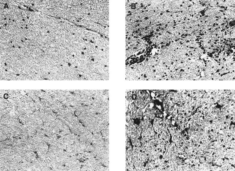
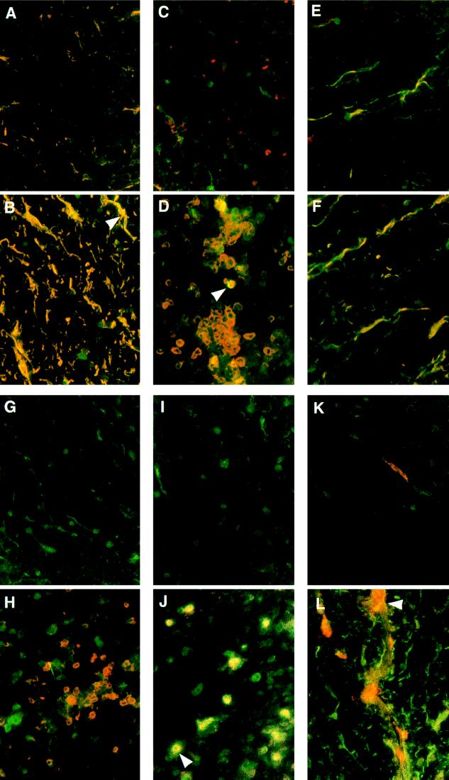
In demyelinating diseases such as multiple sclerosis (MS), myelin membrane structure is destabilized as myelin proteins are lost. Calcium-activated neutral proteinase (calpain) is believed to participate in myelin protein degradation because known calpain substrates [myelin basic protein (MBP); myelin-associated glycoprotein] are degraded in this disease. In exploring the role of calpain in demyelinating diseases, we examined calpain expression in Lewis rats with acute experimental allergic encephalomyelitis (EAE), an animal model for MS. Using double-immunofluorescence labeling to identify cells expressing calpain, we labeled rat spinal cord sections for calpain with a polyclonal millicalpain antibody and with mAbs for glial (GFAP, OX42, GalC) and inflammatory (CD2, ED2, interferon gamma) cell-specific markers. Calpain expression was increased in activated microglia (OX42) and infiltrating macrophages (ED2) compared with controls. Oligodendrocytes (galactocerebroside) and astrocytes (GFAP) had constitutive calpain expression in normal spinal cords whereas reactive astrocytes in spinal cords from animals with EAE exhibited markedly increased calpain levels compared with astrocytes in adjuvant controls. Oligodendrocytes in spinal cords from rats with EAE expressed increased calpain levels in some areas, but overall the increases in calpain expression were small. Most T cells in grade 4 EAE expressed low levels of calpain, but interferon gamma-positive cells demonstrated markedly increased calpain expression. These findings suggest that increased levels of calpain in activated glial and inflammatory cells in EAE may contribute to myelin destruction in demyelinating diseases such as MS.
Figures
References
-
- Martin R, McFarland H F. Crit Rev Clin Lab Sci. 1995;32:121–182. - PubMed
-
- Raine C S. In: Multiple Sclerosis. Hallpike J F, Adams C W, Tourtellotte W W, editors. Baltimore: Williams & Wilkins; 1983. pp. 413–478.
-
- McFarlin D E, McFarland H F. N Eng J Med. 1982;307:1183–1188. - PubMed
-
- McFarlin D E, McFarland H F. N Eng J Med. 1982;307:1246–1251. - PubMed
-
- Hathaway D R, McClelland P. In: Intracellular Calcium-Dependent Proteolysis. Mellgren R L, Murachi T, editors. Boca Raton, FL: CRC; 1990. pp. 91–102.
Publication types
MeSH terms
Substances
Grants and funding
LinkOut - more resources
Full Text Sources
Other Literature Sources
Miscellaneous
