. 1998 May 12;95(10):5779-84.
doi: 10.1073/pnas.95.10.5779.

Neurotrophins inhibit major histocompatibility class II inducibility of microglia: involvement of the p75 neurotrophin receptor

Affiliations

Neurotrophins inhibit major histocompatibility class II inducibility of microglia: involvement of the p75 neurotrophin receptor

H Neumann et al. Proc Natl Acad Sci U S A. .

Abstract

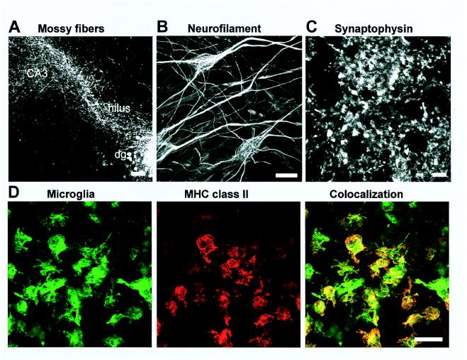
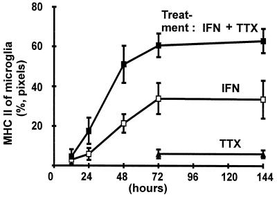
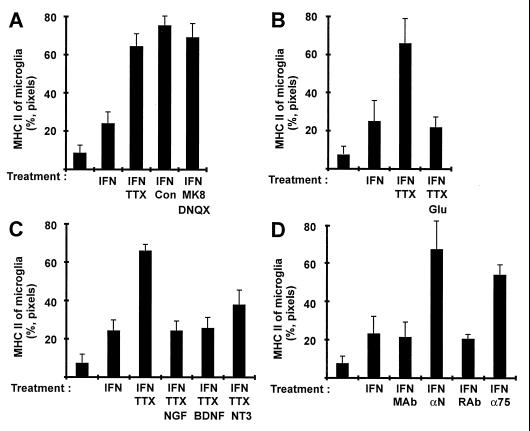
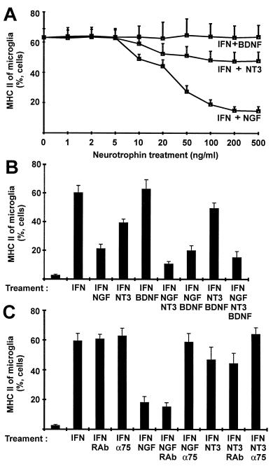
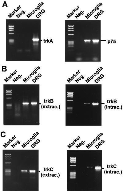
Major histocompatibility complex (MHC) molecules are rare in the healthy brain tissue, but are heavily expressed on microglial cells after inflammatory or neurodegenerative processes. We studied the conditions leading to the induction of MHC class II molecules in microglia by using explant cultures of neonatal rat hippocampus, a model of interacting neuronal networks. Interferon-gamma (IFN-gamma)-dependent MHC class II inducibility in microglia cells was very low, but strongly increased in the hippocampal slices after the blockade of neuronal activity by neurotoxins [tetrodotoxin (TTX), omega-conotoxin] or glutamate antagonists. None of these agents acted directly on isolated microglia cells. We found that neurotrophins modulate microglial MHC class II expression. MHC class II inducibility was enhanced by neutralization of neurotrophins produced locally within the cultured tissues and was inhibited by the addition of nerve growth factor (NGF), brain-derived neurotrophic factor (BDNF), or neurotrophin-3 (NT3). NGF and, to a lower extent, NT3 acted directly on isolated microglia via the p75 neurotrophin receptor and inhibited MHC class II inducibility as shown by blockade of the p75 neurotrophin receptor with antibodies. Our data suggest that neurotrophins secreted by electrically active neurons control the antigen-presenting potential of microglia cells, and indicate that this effect is mediated partly via the p75 neurotrophin receptor.


Figures

Figure 1
Figure 1
Neuronal outgrowth and MHC class II expression of microglia in the cultured hippocampal slices analyzed by confocal laser-scanning microscopy. Neuronal outgrowth was determined by anterograde labeling of mossy fibers innervating the hilus and the CA3 region of the hippocampus (A), by immunolabeling for the cytoskeleton protein neurofilament (B), and by immunolabeling for the synapse protein synaptophysin (C). Slices were treated with IFN-γ (50 units/ml) and double-labeled for MHC class II and isolectin for microglia (D). MHC class II molecules were visualized mainly on microglia. [Bars = 100 μm (A), 20 μm (B), 5 μm (C), 50 μm (D).]
Figure 2
Figure 2
MHC class II inducibility of microglia in the cultured slices as determined by image analysis. Relative expression of MHC class II on microglia (%, pixels) was analyzed by colocalization of pixels on the confocal images. Slices were treated as indicated in the graph for 12, 24, 48, 72, and 144 hr with IFN-γ (IFN, 50 units/ml), IFN-γ plus TTX (IFN, 50 units/ml + TTX, 1 μM), or TTX (TTX, 1 μM) alone. Data are presented as mean and SEM.
Figure 3
Figure 3
Modulation of MHC class II inducibility of microglia in cultured slices by neuronal activity and neurotrophins. Relative expression of MHC class II on microglia (%, pixels) was analyzed on the confocal images by colocalization of pixels. Slices were treated for 72 hr with IFN-γ (IFN, 50 units/ml), and neuronal activity was modulated by TTX (TTX, 1 μM), ω-conotoxin GIVA plus ω-conotoxin MVIIC (Con, 5 μM and 10 μM, respectively), or glutamate antagonist MK801 (MK8, 10 μM) plus DNQX (10 μM) (A). Treatment of the slice cultures with glutamate (20 μM) for 72 hr reversed the TTX-induced up-regulation of MHC class II (B). In addition, NGF (50 ng/ml), BDNF (50 ng/ml), or NT3 (50 ng/ml) was applied for 72 hr and reversed MHC class II expression of microglia in slices treated with IFN-γ and TTX. (C). Slices were treated with IFN-γ (IFN, 50 units/ml), and action of endogenous neurotrophins was inhibited by a mixture of mouse-neutralizing antibodies directed against the neurotrophins NGF/BDNF/NT3 (αN) or by rabbit antibody blocking the p75 neurotrophin receptor (D). As a control slices were treated with equal amounts of total mouse antibody (MAb) or rabbit antibody (RAb), respectively. All data are presented as mean and SEM.
Figure 4
Figure 4
Analysis of MHC class II expression of isolated cultured microglia by flow cytometry. (A) Isolated microglial cells were treated for 72 hr with IFN-γ (IFN, 50 units/ml) and different concentrations of NGF, BDNF, and NT3 for 72 hr. (B) Isolated microglial cells were induced by IFN-γ (IFN, 50 units/ml), and different combinations of NGF (50 ng/ml), BDNF (50 ng/ml), and NT3 (50 ng/ml) were added to the cell cultures for 72 hr. (C) In addition, microglial cells were treated for 72 hr with IFN-γ (IFN, 50 units/ml), and action of NGF (50 ng/ml) and NT3 (50 ng/ml) on MHC class II of microglia was inhibited with rabbit antibody blocking the p75 neurotrophin receptor (α75). Isolated microglial cells were treated with rabbit antibody (RAb) as a control. Data are presented as relative number of cells (mean and SEM).
Figure 5
Figure 5
Analysis of the neurotrophin receptors of isolated microglia (Microglia) and dorsal root ganglia culture (DRG) by RT-PCR. (A) PCR fragments of the trkA receptor were detected in dorsal root ganglia culture, but not in isolated microglia culture. The p75 neurotrophin receptor was detected in both dorsal root ganglia culture and isolated microglial culture. (B and C) PCR fragments of the trkB and trkC receptors were amplified in isolated microglia culture from the extracellular (extrac.) and intracellular (intrac.) domains. The intracellular tyrosine kinase domains of the receptors were detectable at a very low level. Lanes labeled “Marker” and “Neg.” show the molecular weight marker ΦX174/HaeIII-digested and negative PCR control, respectively.

Similar articles

Cited by

References

    1. Wekerle H, Linington C, Lassmann H, Meyermann R. Trends Neurosci. 1986;9:271–277.
    1. Streit W J, Graeber M B, Kreutzberg G W. J Neuroimmunol. 1989;21:117–123. - PMC - PubMed
    1. Itagaki S, McGeer P L, Akiyama H. Neurosci Lett. 1988;91:259–264. - PubMed
    1. Perlmutter L S, Scott S A, Chui H C. J Neurosci Res. 1992;33:549–558. - PubMed
    1. McGeer P L, Itagaki S, McGeer E G. Acta Neuropathol. 1988;76:550–557. - PubMed

Publication types

MeSH terms