. 1998 May 12;95(10):5801-6.
doi: 10.1073/pnas.95.10.5801.

GFRalpha3 is an orphan member of the GDNF/neurturin/persephin receptor family

Affiliations

GFRalpha3 is an orphan member of the GDNF/neurturin/persephin receptor family

R H Baloh et al. Proc Natl Acad Sci U S A. .

Abstract

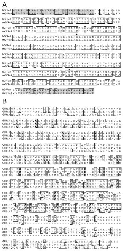
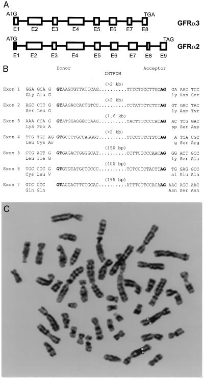
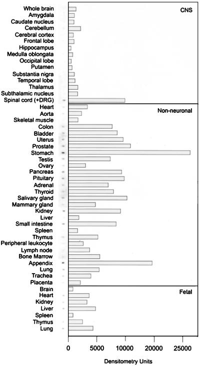
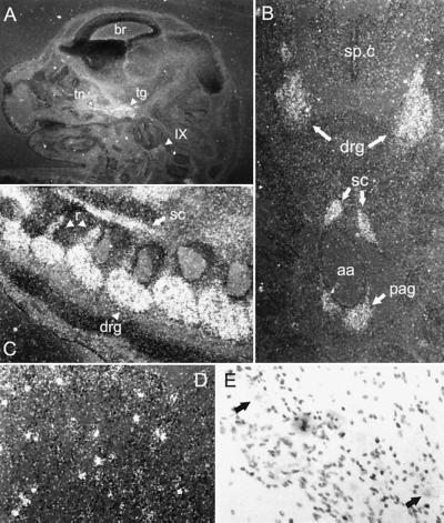
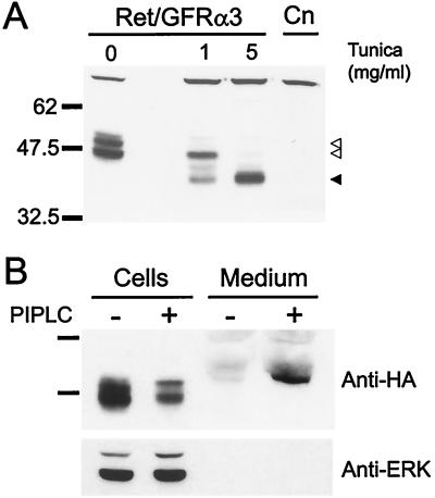
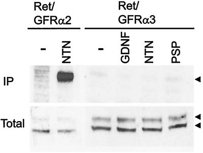
GDNF, neurturin, and persephin are transforming growth factor beta-related neurotrophic factors known collectively as the GDNF family (GF). GDNF and neurturin signal through a multicomponent receptor complex containing a signaling component (the Ret receptor tyrosine kinase) and either of two glycosyl-phosphatidylinositol-linked binding components (GDNF family receptor alpha components 1 and 2, GFRalpha1 or GFRalpha2), whereas the receptor for persephin is unknown. Herein we describe a third member of the GF coreceptor family called GFRalpha3 that is encoded by a gene located on human chromosome 5q31.2-32. GFRalpha3 is not expressed in the central nervous system of the developing or adult animal but is highly expressed in several developing and adult sensory and sympathetic ganglia of the peripheral nervous system. GFRalpha3 is also expressed at high levels in developing, but not adult, peripheral nerve. GFRalpha3 is a glycoprotein that is glycosyl-phosphatidylinositol-linked to the cell surface like GFRalpha1 and GFRalpha2. Fibroblasts expressing Ret and GFRalpha3 do not respond to any of the known members of the GDNF family, suggesting that GFRalpha3 interacts with an unknown ligand or requires a different or additional signaling protein to function.


Figures

Figure 1
Figure 1
Sequence analysis and alignment of GFRα3. (A) Alignment of the amino acid sequences of human and mouse GFRα3. Identical residues are boxed, the N- and C-terminal pro regions are shaded, and putative N-linked glycosylation sites are marked by dots. (B) Alignment of mouse GFRα family members. Identical residues are boxed, and shared cysteines are shaded. Note the divergence of GFRα3 sequence, particularly the large deletion in the C-terminal region.
Figure 2
Figure 2
Genomic analysis of GFRα3. (A) Intron–exon junctions in the coding region of GFRα2 and GFRα3. Exons are represented by boxes and scaled according to size. Introns (intervening lines) are not to scale. Note that GFRα3 entirely lacks one exon relative to GFRα2. (B) Precise intron sites in the mouse GFRα3 gene. Consensus splice sites are shown in boldface type. (C) Fluorescence in situ hybridization analysis of the human GFRα3 gene location. Digital image of a metaphase chromosome spread derived from methotrexate-synchronized normal human peripheral leukocytes after hybridization with a digoxigenin-labeled GFRα3 genomic probe and 4′,6-diamidino-2-phenylindole counterstaining. Both copies of chromosome 5 have symmetrical rhodamine signals on sister chromatids at region 5q 31.2–32.
Figure 3
Figure 3
Survey of GFRα3 expression in human tissues. A human MRB (CLONTECH) was probed with a fragment of the human GFRα3 cDNA. The hybridization signals are shown next to the signal intensity quantified by using a PhosphorImager. GFRα3 was low or absent in structures of the CNS and moderate in some nonneuronal tissues, particularly those of the digestive system, urogenital system, and several glandular structures. Note that the spinal cord sample contained DRG material, which is likely the source of GFRα3 expression (see text and Fig. 4).
Figure 4
Figure 4
In situ hybridization analysis of GFRα3 expression in mouse. (A) Saggital section of E14 mouse head. GFRα3 is observed in trigeminal ganglion (tg) and nerve (tn) and in the glossopharyngeal ganglion (IX) but not in brain (br). (B) Transverse section of E14 mouse showing GFRα3 expression in DRG but not in the spinal cord (sp.c). Around the abdominal aorta (aa), staining is observed in the sympathetic chain ganglia (sc) and the preaortic ganglia of Zuckerkandl (pag). (C) Saggital section of E14 mouse spinal column showing DRGs (drg), sympathetic chain (sc), and labeled nerve roots (r). (D) Dark-field photomicrograph of adult trigeminal ganglion showing punctate staining in contrast to the diffuse staining observed at E14. (E) Bright-field photomicrograph of adult trigeminal ganglion at higher power, showing GFRα3 is localized to neurons (arrows).
Figure 5
Figure 5
Analysis of NHA-GFRα3 protein in transfected fibroblasts. (A) Anti-HA immunoblot of fibroblasts stably transfected with HA-tagged GFRα3 and Ret (Ret/GFRα3) show a doublet at approximately 47 and 51 kDa (open arrowheads) that was not present in the parental line (Cn). Tunicamycin (Tunica) treatment of the cells for 24 hr to block N-linked glycosylation resulted in loss of the upper band at 1 mg/ml, and appearance of a lower molecular mass band. At a tunicamycin concentration of 5 mg/ml, only the lower band running at the predicted molecular mass for GFRα3 was visible (solid arrowhead). (B) Phosphatidylinositol-specific phospholipase C (PIPLC) treatment of NHA-GFRα3 expressing fibroblasts to specifically cleave GPI-linked proteins resulted in depletion of the NHA-GFRα3 from the cells and induced the presence of a band in the medium corresponding to NHA-GFRα3. Reprobing of the blot with an anti-ERK p42/44 antibody is shown below to indicate equal loading of cell lysates.
Figure 6
Figure 6
Functional analysis of the fibroblasts expressing Ret and GFRα3. Fibroblasts that stably express Ret and GFRα2 (Ret/GFRα2) or Ret and NHA-GFRα3 (Ret/GFRα3) were left untreated (−) or stimulated with the indicated GF ligand (100 ng/ml) for 10 min, immunoprecipitated (IP) with anti-phosphotyrosine antibodies, separated by SDS/PAGE, and immunoblotted with an anti-Ret antibody. Fractions of the total lysates before immunoprecipitation are shown below to demonstrate presence of Ret (arrowheads) and equal loading (Total). As expected, stimulation of Ret/GFRα2 containing fibroblasts with NTN shows strong Ret phosphorylation, whereas stimulation of Ret/GFRα3-containing fibroblasts with GDNF, NTN, or PSP did not lead to Ret phosphorylation, indicating GFRα3 is not able to form a functional receptor with Ret for any of these GF ligands.

Similar articles

Cited by

References

    1. Lin L-F H, Doherty D H, Lile J D, Bektesh S, Collins F. Science. 1993;260:1130–1132. - PubMed
    1. Kotzbauer P T, Lampe P A, Heuckeroth R O, Golden J P, Creedon D J, Johnson E M, Milbrandt J D. Nature (London) 1996;384:467–470. - PubMed
    1. Milbrandt J, de Sauvage F, Fahrner T J, Baloh R H, Leitner M L, Tansey M G, Lampe P A, Heuckeroth R O, Kotzbauer P T, Simburger K S, et al. Neuron. 1998;20:245–253. - PubMed
    1. Stromberg I, Bjorklund L, Johansson M, Tomac A, Collins F, Olson L, Hoffer B, Humpel C. Exp Neurol. 1993;124:401–412. - PubMed
    1. Hudson J, Granholm A-C, Gerhardt G A, Henry M A, Hoffman A, Biddle P, Leela N S, Mackerlova L, Lile J D, Collins F, Hoffer B J. Brain Res Bull. 1995;36:425–432. - PubMed

Publication types

MeSH terms

Associated data