. 1998 May;8(5):549-56.
doi: 10.1101/gr.8.5.549.

A homogeneous, ligase-mediated DNA diagnostic test

Affiliations

A homogeneous, ligase-mediated DNA diagnostic test

X Chen et al. Genome Res. 1998 May.

Abstract

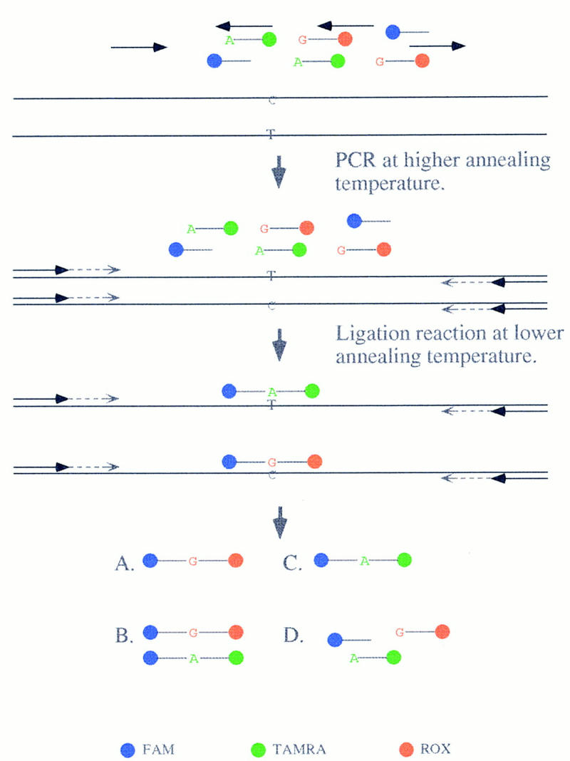
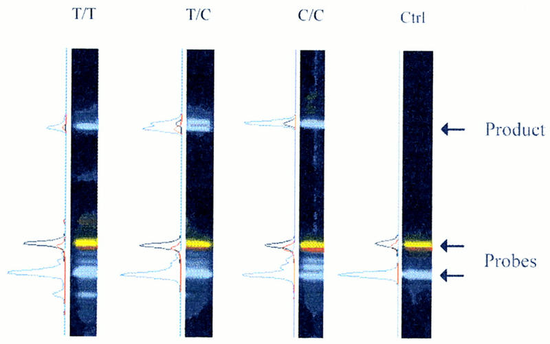
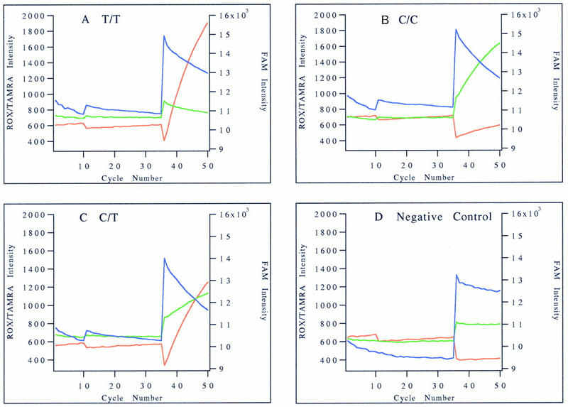
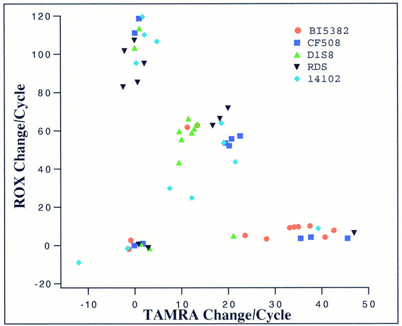
Single-nucleotide variations are the most widely distributed genetic markers in the human genome. A subset of these variations, the substitution mutations, are responsible for most genetic disorders. As single nucleotide polymorphism (SNP) markers are being developed for molecular diagnosis of genetic disorders and large-scale population studies for genetic analysis of complex traits, a simple, sensitive, and specific test for single nucleotide changes is highly desirable. In this report we describe the development of a homogeneous DNA detection method that requires no further manipulations after the initial reaction is set up. This assay, named dye-labeled oligonucleotide ligation (DOL), combines the PCR and the oligonucleotide ligation reaction in a two-stage thermal cycling sequence with fluorescence resonance energy transfer (FRET) detection monitored in real time. Because FRET occurs only when the donor and acceptor dyes are in close proximity, one can infer the genotype or mutational status of a DNA sample by monitoring the specific ligation of dye-labeled oligonucleotide probes. We have successfully applied the DOL assay to genotype 10 SNPs or mutations. By designing the PCR primers and ligation probes in a consistent manner, multiple assays can be done under the same thermal cycling conditions. The standardized design and execution of the DOL assay means that it can be automated for high-throughput genotyping in large-scale population studies.


Figures

Figure 1
Figure 1
DOL assay with FRET detection. All of the reagents, including DNA polymerase, DNA ligase, PCR primers, and ligation probes, are added to the test DNA sample when the reaction is first set up. In a two-stage thermal cycling sequence, PCR is allowed to proceed without the ligation probe annealing to the target followed by the ligation reaction when enough PCR product has accumulated. At the end of the assay, there are four possible outcomes for each test sample: (A) homozygous for the first allele leading to the formation of a ligation product containing the G allele in this example in the absence of the ligation product corresponding to the second allele (A allele in this example); (B) heterozygous with ligation product of both alleles formed; (C) homozygous for the second allele with the ligation product containing the A allele formed in this example in the absence of the G allele ligation product; (D) PCR or ligation failure leading to no ligation product being formed. (Blue) FAM; (green) TAMRA; (red) ROX.
Figure 2
Figure 2
Gel image and electropherograms of PCR–DOL reaction mixtures. Four DNA samples were subjected to the PCR–DOL assay to test their mutational status for the C/T βo-thalassemia mutation. The unreacted ligation probes are found at the bottom of the gel image [(blue band) FAM-labeled common probe; (red band) ROX-labeled mutant T probe; (yellow band) TAMRA-labeled wild-type C probe]. The products are found near the top of the gel image, where the FAM–ROX ligation product migrates slightly faster than the FAM–TAMRA ligation product. The electropherogram of each lane is plotted alongside the gel image where the color scheme is the same as that for the gel image, except that the black peak represents the TAMRA label. (Lane 1) Homozygous mutant sample with a single product that is doubly labeled with FAM and ROX. (Lane 2) Heterozygous carrier with two products that are doubly labeled with FAM–TAMRA and FAM–ROX, respectively. (Lane 3) Homozygous normal control with a single product doubly labeled with FAM and TAMRA. Varying amounts of FAM-probe extension products (blue bands between the unreacted FAM and ROX/TAMRA probes) are seen in lanes 1–3. (Lane 4) Negative control salmon sperm DNA sample in which no ligation or extension products are seen.
Figure 3
Figure 3
Fluorescence intensity profiles of the PCR–DOL assay. The real-time fluorescence intensities of FAM (blue), ROX (red), and TAMRA (green) during PCR and ligation phases of the reaction are plotted against cycle number. The scale for ROX and TAMRA intensities is on the left, and that for FAM intensity is on the right. The fluorescence intensity profiles of the same samples used for Fig. 2 are shown here. (A) Homozygous mutant sample; (B) homozygous normal control; (C) heterozygous carrier; (D) negative salmon sperm DNA control.
Figure 4
Figure 4
Scatter plot of initial slopes of change in fluorescence intensities. The linearly normalized initial slopes of ROX and TAMRA fluorescence intensity changes of 60 samples (10 human genomic plus 2 salmon sperm DNA samples for each of the 5 markers/mutations) are plotted together. A normalization constant is used for each marker such that the data points can be plotted together on the same scale.

References

    1. Chen X, Kwok P-Y. Template-directed dye-terminator incorporation (TDI) assay: A homogeneous DNA diagnostic method based on fluorescence energy transfer. Nucleic Acids Res. 1997;25:347–353. - PMC - PubMed
    1. Chen X, Zehnbauer B, Gnirke A, Kwok P-Y. Fluorescence energy transfer detection as a homogeneous DNA diagnostic method. Proc Natl Acad Sci. 1997;94:10756–10761. - PMC - PubMed
    1. Collins FS, Guyer MS, Chakravarti A. Variations on a theme: Cataloging human DNA sequence variation. Science. 1997;278:1580–1581. - PubMed
    1. Couch FJ, Weber BL. Mutations and polymorphisms in the familial early-onset breast cancer (BRCA1) gene. Hum Mutat. 1996;8:8–18. - PubMed
    1. Eggerding FA. A one-step coupled amplification and oligonucleotide ligation procedure for multiplex genetic typing. PCR Methods Applic. 1995;4:337–345. - PubMed

Publication types