. 1998 Jun;18(6):3149-62.
doi: 10.1128/MCB.18.6.3149.

The regulatory particle of the Saccharomyces cerevisiae proteasome

Affiliations

The regulatory particle of the Saccharomyces cerevisiae proteasome

M H Glickman et al. Mol Cell Biol. 1998 Jun.

Abstract

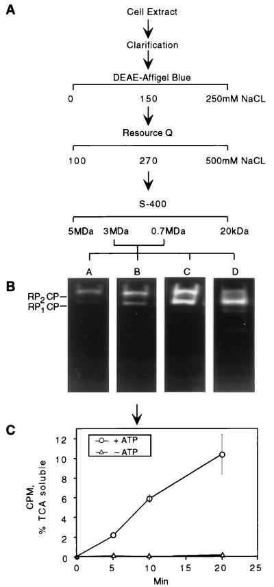
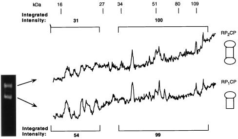
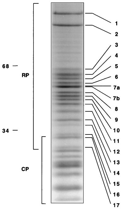
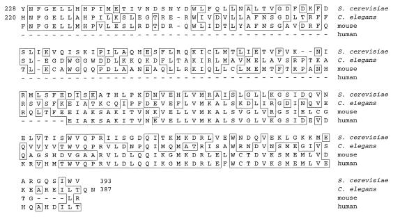
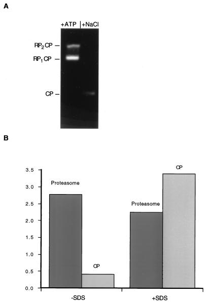
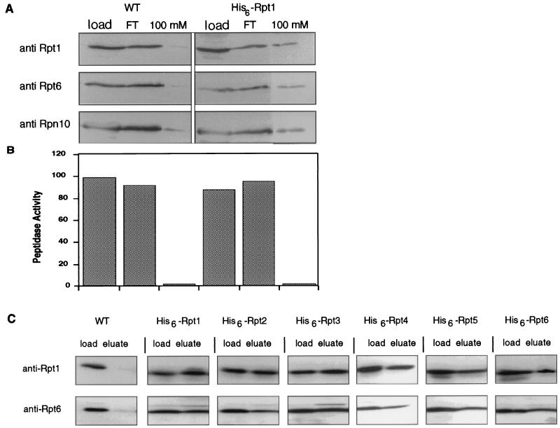
The proteasome is a multisubunit protease responsible for degrading proteins conjugated to ubiquitin. The 670-kDa core particle of the proteasome contains the proteolytic active sites, which face an interior chamber within the particle and are thus protected from the cytoplasm. The entry of substrates into this chamber is thought to be governed by the regulatory particle of the proteasome, which covers the presumed channels leading into the interior of the core particle. We have resolved native yeast proteasomes into two electrophoretic variants and have shown that these represent core particles capped with one or two regulatory particles. To determine the subunit composition of the regulatory particle, yeast proteasomes were purified and analyzed by gradient sodium dodecyl sulfate-polyacrylamide gel electrophoresis. Resolution of the individual polypeptides revealed 17 distinct proteins, whose identities were determined by amino acid sequence analysis. Six of the subunits have sequence features of ATPases (Rpt1 to Rpt6). Affinity chromatography was used to purify regulatory particles from various strains, each of which expressed one of the ATPases tagged with hexahistidine. In all cases, multiple untagged ATPases copurified, indicating that the ATPases assembled together into a heteromeric complex. Of the remaining 11 subunits that we have identified (Rpn1 to Rpn3 and Rpn5 to Rpn12), 8 are encoded by previously described genes and 3 are encoded by genes not previously characterized for yeasts. One of the previously unidentified subunits exhibits limited sequence similarity with deubiquitinating enzymes. Overall, regulatory particles from yeasts and mammals are remarkably similar, suggesting that the specific mechanistic features of the proteasome have been closely conserved over the course of evolution.


Figures

FIG. 1
FIG. 1
Proteasome purification procedure. (A) A yeast lysate was fractionated on a series of columns containing DEAE–Affi-Gel Blue, Resource Q resin, or S-400 resin (see Materials and Methods for details). Fractions containing peptidase activity were combined into four pools (A to D) in descending molecular mass order. Protein content and specific peptidase activity at each step are shown in Table 1. (B) Proteasomes from each pool were visualized by nondenaturing PAGE and fluorogenic peptide overlay. In pool D, two faster-migrating species were observed in addition to RP2CP and RP1CP. The fastest-migrating species was the CP, and the other contained the CP and a subset of RP subunits (data not shown). (C) Proteasomes from pool B were tested for the ability to proteolyse multiubiquitinated 125I-labeled lysozyme in the presence or absence of ATP. Degradation was measured as trichloroacetic acid-soluble 125I counts per minute at a given time point. Background radioactivity was subtracted from all readings. Error bars indicate standard deviations.
FIG. 2
FIG. 2
The proteasome migrates as singly and doubly capped forms on nondenaturing PAGE. Purified proteasomes from pool B were resolved by nondenaturing PAGE with a reversible cross-linker, N,N′-bisacrylylcystamine. The fast- and slow-migrating forms were excised. Their proteins were extracted from the gels and resolved by SDS–12% PAGE. The gels were then stained with Coomassie blue. Densitometric quantitation of the resulting protein banding pattern is shown for each form. The protein banding pattern can be compared to that shown in Fig. 3 prior to nondenaturing PAGE. However, as the two gel systems are different, the comparison cannot be made on a one-to-one basis. Based on the data in Table 2 (and data not shown), proteins below 25 kDa were assumed to be CP subunits, and those above 30 kDa were assigned as be RP subunits. The integrated intensities of CP and RP subunits are displayed over the corresponding region of the gel. The ratio of intensity of RP subunits to that of CP subunits in the slower-migrating form of the proteasome is approximately double that in the faster-migrating form.
FIG. 3
FIG. 3
Subunit composition of the proteasome determined by gradient SDS-PAGE. Proteins from pool B (Fig. 1) were resolved on a 10 to 20% polyacrylamide gradient gel. Protein bands were stained with Coomassie blue. Seventeen protein bands in the 120- to 30-kDa region were numbered in descending molecular mass order (masses are shown on the left). Proteins were excised from the gel and digested with trypsin. The resulting peptides were separated by reverse-phase HPLC and subjected to sequence analysis. Peptides sequenced from each band are shown in Table 2.
FIG. 4
FIG. 4
Rpn9 is homologous to putative proteasome subunits in other eukaryotes. Homology of the C terminus of Rpn9 to deduced protein sequences of the product of a hypothetical ORF in C. elegans (z49130) and mouse (w34049) and human (aa122133) EST fragments is shown. The alignment was obtained by the Jotun Hein method with MegAlign (gap penalty, 11; gap length, 3). The C terminus of Rpn9 was 35, 36, and 42% identical to the C. elegans, mouse, and human sequences, respectively. Boxes indicate amino acid identities; dashes indicate gaps in the alignment.
FIG. 5
FIG. 5
Temperature-sensitive phenotype caused by Δrpn9 deletion mutation. (A) Wild-type (SUB62), myc6-RPN9 (MG26), and Δrpn9 (MG18) strains were grown on YPD at 30°C (top panel) or 37°C (bottom panel). The Δrpn9 strain was temperature sensitive, showing no detectable growth after 48 h at 37°C. (After 1 week, a few small colonies were observed.) The myc6-tagged version of RPN9 fully complemented the deletion. (B and C) The Δrpn9Δrpn10 double mutant did not display a marked synthetic phenotype. Wild-type (SUB62), Δrpn9 (MG18), Δrpn10 (102), and Δrpn9Δrpn10 double mutant (MG29) strains were grown for 48 h on YPD at either 30°C (B) or 37°C (C).
FIG. 6
FIG. 6
Rpn9 is an RP subunit. Proteasomes from wild-type (wt) and myc6-RPN9 strains were partially purified on a DEAE–Affi-Gel Blue column and further resolved by nondenaturing PAGE. (A) Proteasome bands visualized in situ by peptidase activity against Suc-LLVY-AMC. (B and C) Immunoblots probed with the indicated antibodies.
FIG. 7
FIG. 7
Structural alignment of six proteasomal ATPases. Comparison of the six Rpt subunits based on their primary structure shows a highly conserved ATPase module (black box) containing the A and B loops which form the predicted ATP binding domain (33, 34, 67, 103). The N termini are variable, in some genes containing a predicted (62) coiled-coil domain (open box).
FIG. 8
FIG. 8
The proteasome is a heteromeric complex of ATPases. His6-Rpt2 was expressed in a Δrpt2 background (DY17). Extracts from His6-Rpt2-expressing and wild-type (WT) control strains were partially purified by DEAE–Affi-Gel Blue chromatography in the presence of 1 mM Mg-ATP. The 150 mM NaCl eluate was subjected to Ni-NTA affinity chromatography. Column fractions were immunoblotted (A) and tested for peptidase activity against Suc-LLVY-AMC (B). The epitope-tagged complex eluted at 100 mM imidazole, as indicated by immunoblotting against Rpt1, Rpt6, and Rpn10 (A) and by peptidase activity (B). The wild-type proteasome eluted during low-imidazole rinses. (C) Extracts from strains expressing His6-tagged versions of each of the six ATPases were individually purified by Ni-NTA chromatography. Fractions loaded onto the Ni-NTA column (Load) were compared to fractions from the 100 mM imidazole eluate (Eluate) by immunoblotting with anti-Rpt1 and anti-Rpt6 antibodies.
FIG. 9
FIG. 9
Dissociation of the RP from the CP inhibits peptidase activity. Equal amounts of purified proteasome were incubated for 30 min at 30°C in buffer A or in buffer A without Mg-ATP but with 500 mM NaCl. (A) The two samples were resolved by nondenaturing PAGE and visualized by activity against the fluorogenic peptide substrate Suc-LLVY-AMC. After incubation in 500 mM NaCl, both singly- and doubly-capped forms of the proteasome (RP2CP and RP1CP, respectively; left lane) disassembled, giving rise to free CPs (right lane). It is apparent that the CP had lower peptidase activity than the proteasome on a molar level. The RP was not visualized by this method, as it contains no intrinsic peptidase activity. (B) To quantify the difference in peptidase activities between the proteasome and the CP, approximately equimolar quantities of the two samples were incubated for 10 min at 30°C in buffer A with 0.1 mM fluorogenic peptide Suc-LLVY-AMC; the fluorescence of released AMC is shown in the left columns. The two samples were also incubated for 10 min at 30°C in buffer A with 0.1 mM Suc-LLVY-AMC and 0.02% SDS (right columns). The CP exhibited a lower level of peptidase activity and a higher level of SDS stimulation than the intact proteasome.
FIG. 10
FIG. 10
Proteasomal ATPases associate into a heteromeric complex. His6-Rpt1 was expressed in a Δrpt1 background (DY19). Extracts from His6-Rpt1-expressing and wild-type (WT) control strains were partially purified on DEAE–CL-6B resin in the absence of ATP. The 500 mM NaCl eluate was fractionated on Ni-NTA affinity columns. Column fractions were subjected to immunoblotting (A) and tested for peptidase activity against Suc-LLVY-AMC (B). The epitope-tagged complex eluting at 100 mM imidazole contained a number of RP subunits (Rpt1, Rpt6, and Rpn10) (A) but lacked peptidase activity (B). The wild-type complex eluted during low-imidazole rinses. (C) Extracts from strains expressing His6-tagged versions of each of the six ATPases were also purified by Ni-NTA chromatography. Fractions loaded onto the Ni-NTA column (Load) were compared to fractions from the 100 mM imidazole eluate (Eluate) by immunoblotting with anti-Rpt1 and anti-Rpt6 antibodies.

References

    1. Adams G M, Falke S, Goldberg A L, Slaughter C A, DeMartino G N, Gogol E P. Structural and functional effects of PA700 and modulator protein on proteasomes. J Mol Biol. 1997;273:646–657. - PubMed
    1. Akiyama K, Yokota K, Kagawa S, Shimbara N, DeMartino G N, Slaughter C A, Noda C, Tanaka K. cDNA cloning of a new putative ATPase subunit p45 of the human 26S proteasome, a homolog of yeast Sug1. FEBS Lett. 1995;363:151–156. - PubMed
    1. Armon T, Ganoth D, Hershko A. Assembly of the 26S complex that degrades proteins ligated to ubiquitin is accompanied by the formation of ATPase activity. J Biol Chem. 1990;265:20723–20726. - PubMed
    1. Baker R T, Tobias J W, Varshavsky A. Ubiquitin-specific proteases of S. cerevisiae. Cloning of UBP2 and UBP3 and functional analysis of the UBPgene family. J Biol Chem. 1992;267:23364–23375. - PubMed
    1. Bauer V W, Swaffield J C, Johnston S A, Andrews M T. CADp44: a novel regulatory subunit of the 26S proteasome and the mammalian homolog of ySug2p. Gene. 1996;181:63–69. - PubMed

Publication types

MeSH terms