. 1998 Jun;18(6):3245-56.
doi: 10.1128/MCB.18.6.3245.

RACK1, a receptor for activated C kinase and a homolog of the beta subunit of G proteins, inhibits activity of src tyrosine kinases and growth of NIH 3T3 cells

Affiliations

RACK1, a receptor for activated C kinase and a homolog of the beta subunit of G proteins, inhibits activity of src tyrosine kinases and growth of NIH 3T3 cells

B Y Chang et al. Mol Cell Biol. 1998 Jun.

Abstract

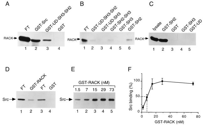
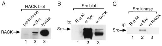
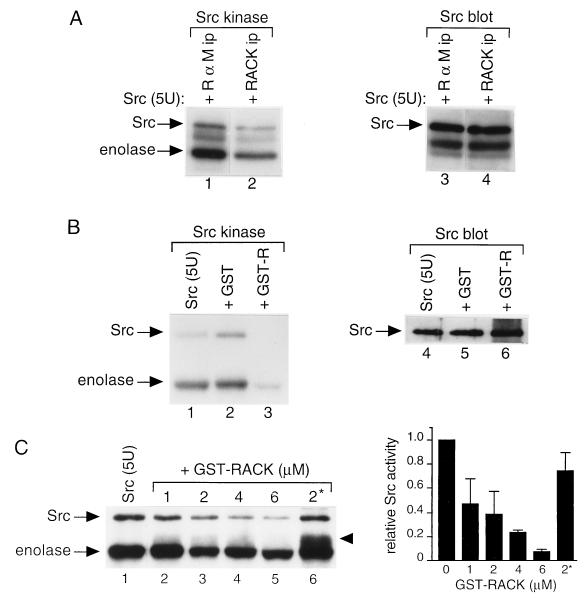
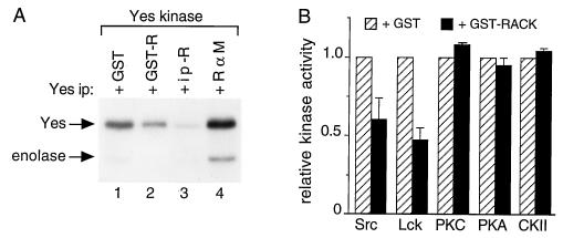
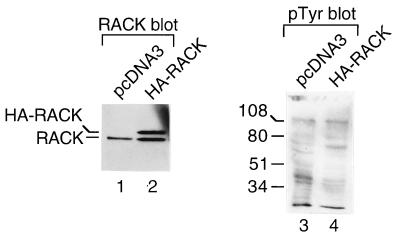
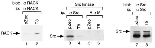
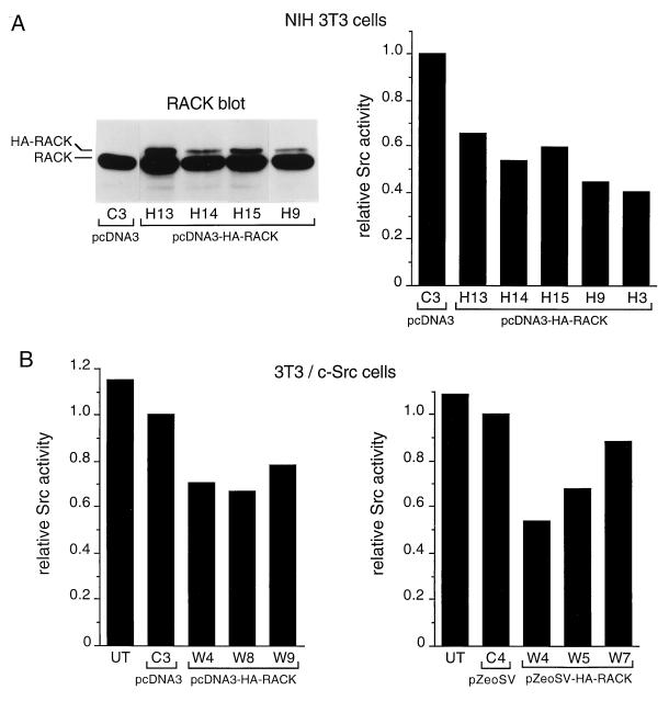
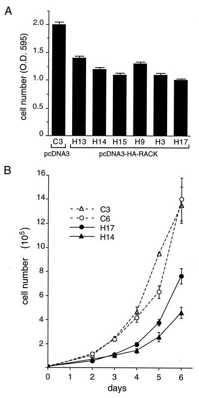
To isolate and characterize proteins that interact with the unique domain and SH3 and SH2 domains of Src and potentially regulate Src activity, we used the yeast two-hybrid assay to screen a human lung fibroblast cDNA library. We identified RACK1, a receptor for activated C kinase and a homolog of the beta subunit of G proteins, as a Src-binding protein. Using GST-Src fusion proteins, we determined that RACK1 binds to the SH2 domain of Src. Coimmunoprecipitation of Src and RACK1 was demonstrated with NIH 3T3 cells. Purified GST-RACK1 inhibited the in vitro kinase activity of Src in a concentration-dependent manner. GST-RACK1 (2 microM) inhibited the activities of purified Src and Lck tyrosine kinases by 40 to 50% but did not inhibit the activities of three serine/threonine kinases that we tested. Tyrosine phosphorylation on many cellular proteins decreased in 293T cells that transiently overexpressed RACK1. Src activity and cell growth rates decreased by 40 to 50% in NIH 3T3 cells that stably overexpressed RACK1. Flow cytometric analyses revealed that RACK1-overexpressing cells do not show an increased rate of necrosis or apoptosis but do spend significantly more time in G0/G1 than do wild-type cells. Prolongation of G0/G1 could account for the increased doubling time of RACK1-overexpressing cells. We suggest that RACK1 exerts its effect on the NIH 3T3 cell cycle in part by inhibiting Src activity.


Figures

FIG. 1
FIG. 1
Association of RACK1 and Src in vitro. (A) Binding of in vitro-translated RACK1 to GST-Src fusion proteins. [35S]methionine- and [35S]cysteine-labeled RACK1 was synthesized in rabbit reticulocyte lysates and incubated with 1 μg of purified GST-Src (lane 2), GST-UD/SH3/SH2 (lane 3), or GST (lane 4) for 3 h at 4°C. Protein complexes were collected on glutathione-agarose beads, washed, and boiled in SDS sample buffer. Proteins were resolved by SDS-PAGE. For each reaction, 1/10 of the unbound translation reaction product (flowthrough [FT]) was loaded directly on the gel as a measure of the amount of protein translated (lane 1). 35S-labeled proteins were visualized by autoradiography and quantified by scanning densitometry. (B) Binding of in vitro-translated RACK1 to additional GST-Src fusion proteins. In vitro-translated RACK1 (lane 1) was assessed for binding to GST-UD/SH3/SH2 (lane 2), GST-UD/SH3 (lane 3), GST-SH2/SH3 (lane 4), GST-SH3 (lane 5), or GST-SH2 (lane 6) as described for panel A. (C) Binding of RACK1 from HeLa cell lysates to GST-Src fusion proteins. HeLa cell lysates containing 500 μg of total cellular protein were incubated with 5 μg of GST-SH2 (lane 2), GST alone (lane 3), GST-SH3 (lane 4), or GST-UD (lane 5) for 3 h at 4°C. Proteins bound to GST or GST-Src fusion proteins were recovered, resolved by SDS-PAGE, transferred to PVDF membranes, and subjected to immunoblot analysis with anti-RACK1 MAb. Lysate containing 20 μg of total cellular protein was loaded directly on the gel prior to transfer and immunoblotting with anti-RACK1 MAb (lane 1). (D) Binding of in vitro-translated Src to GST-RACK1. In vitro-translated Src was assessed for binding to GST-RACK1 (lane 2) or GST alone (lane 4) as described in panel A. For each reaction, 1/20 of the FT was loaded directly on the gel (lanes 1 and 3). (E) Concentration-dependent binding of GST-RACK1 to in vitro-translated Src. A constant amount of in vitro-translated Src was assessed for binding to increasing concentrations of GST-RACK1 (1.5 to 73 nM). Data are representative of four independent experiments. (F) Quantitation of data on concentration-dependent binding of GST-RACK1 to in vitro-translated Src. Data represent average values from four independent experiments and are expressed as a percentage of maximal Src binding. Error bars indicate standard errors. Broken lines show the amount of GST-RACK1 required for half-maximal Src binding.
FIG. 2
FIG. 2
Association of RACK1 and Src in vivo. (A) RACK1 coimmunoprecipitates with Src. Proteins were precipitated with preimmune serum (lane 1) or excess Src polyclonal antibody R7 (lane 2) from NP-40 lysates of NIH 3T3 cells containing 800 μg of total cellular protein, resolved by SDS-PAGE, transferred to PVDF membranes, and immunoblotted with anti-RACK1 antibody. Lysate containing 20 μg of total cellular protein was loaded directly on the gel prior to transfer and immunoblot analysis with anti-RACK1 antibody (lane 3). (B) Src coimmunoprecipitates with RACK1. Proteins were immunoprecipitated (ip) with rabbit anti-mouse IgG (R α M, lane 1), Src MAb 327 (lane 2), or RACK1 MAb (lane 3) from NP-40 lysates of 3T3/c-Src cells containing 500 μg of total cellular protein and subjected to immunoblot analysis with Src MAb 327. The band below Src is mouse IgG heavy chain. (C) In vitro protein kinase activity of Src bound to RACK1. Immunoprecipitates parallel to those shown in panel B were incubated with [γ-32P]ATP and MnCl2 for 10 min at 30°C in an in vitro protein kinase assay. 32P-labeled proteins were resolved by SDS-PAGE and visualized by autoradiography.
FIG. 3
FIG. 3
Effect of RACK1 on Src activity in vitro. (A) Effect of RACK1 immunoprecipitates on the activity of purified Src. Proteins were immunoprecipitated (ip) with excess anti-RACK1 antibody (lanes 2 and 4) or rabbit anti-mouse IgG (lanes 1 and 3) from NP-40 lysates of NIH 3T3 cells containing 100 μg of total cellular protein, incubated with 5 U of purified Src, and subjected to an in vitro protein kinase assay (lanes 1 and 2) or immunoblot analysis with Src MAb 327 (lanes 3 and 4). The band below Src is IgG heavy chain. (B) Effect of purified GST-RACK1 on the activity of purified Src. In vitro protein kinase assays (lanes 1 and 3) or immunoblot analysis with MAb 327 (lanes 4 to 6) was performed with 5 U of purified Src alone (lanes 1 and 4) or with the addition of 2 μM GST (lanes 2 and 5) or GST-RACK1 (GST-R) (lanes 3 and 6). (C) Concentration-dependent inhibition of Src activity by GST-RACK1. (Left panel) In vitro protein kinase assays were performed with 5 U of purified Src together with 2 μM GST (lane 1), 1 to 6 μM GST-RACK1 (lanes 2 to 5), or 2 μM boiled GST-RACK1 (2*) (lane 6). 32P-labeled proteins were resolved by SDS-PAGE and visualized by autoradiography. The arrowhead indicates a 55-kDa phosphorylated protein that comigrated with Coomassie-blue stained GST-RACK1. Data are representative of four independent experiments. (Right panel) Quantitation of data on concentration-dependent inhibition of Src activity by GST-RACK1. 32P incorporation into Src was quantified by scanning densitometry. Data represent average values from four independent experiments and are expressed relative to those for purified Src with the addition of GST alone. Error bars indicate standard errors.
FIG. 4
FIG. 4
Effect of RACK1 on the activities of Src family and serine/threonine protein kinases. (A) Effect of RACK1 on Yes kinase activity. Proteins were precipitated with excess antipeptide antibody specific for Yes from RIPA lysates of WiDr colon carcinoma cells containing 500 μg of total cellular protein. Yes immunoprecipitates were incubated with 2 μM GST (lane 1) or GST-RACK1 (GST-R) (lane 2) or with anti-RACK1 antibody (R) (lane 3) or rabbit anti-mouse (R α M) IgG (lane 4) immunoprecipitates (ip) of WiDr cell lysates containing 100 μg of total cellular protein for 15 min at 4°C and subjected to an in vitro kinase assay with enolase as an exogenous substrate. (B) Effect of GST-RACK1 on the activities of Src family and serine/threonine protein kinases. Purified kinases were incubated with 2 μM GST or GST-RACK1 and assayed for in vitro protein kinase activity with specific peptides as substrates. All assays were performed with equivalent specific activities for each kinase. Data represent average values from three independent measurements of Src or Lck activity and two independent measurements of PKC, PKA, or CKII activity. Data on kinase activity measured after the addition of GST-RACK1 (solid bars) are expressed relative to those measured after the addition of GST alone (hatched bars). Error bars indicate standard errors.
FIG. 5
FIG. 5
Effect of RACK1 overexpression on protein tyrosine phosphorylation in vivo. 293T cells were transiently transfected with 1 μg of pcDNA3 (lanes 1 and 3) or pcDNA3-HA-RACK1 (lanes 2 and 4). After 30 h, cells were treated with 100 μM sodium vanadate for 30 min and lysed. Lysates were resolved by SDS-PAGE, transferred to PVDF membranes, and incubated with anti-RACK1 antibody (lanes 1 and 2) or antiphosphotyrosine antibody PY20 (pTyr) (lanes 3 and 4). Protein sizes are indicated in kilodaltons.
FIG. 6
FIG. 6
Effect of RACK1 overexpression on Src activity. NIH 3T3 cells were transfected with pZeoSV (pZeo) or pZeoSV-RACK1, and isolated clones that stably overexpressed RACK1 were assayed for Src kinase activity. The results from one clone, T8, are shown. Proteins were immunoprecipitated (ip) from lysates containing 500 μg of total cellular protein with excess anti-RACK1 MAb (lanes 1 and 2), Src MAb 327 (lanes 3, 4, 7, and 8) or rabbit anti-mouse (R α M) IgG (lanes 5 and 6) and subjected to immunoblot analysis with anti-RACK1 MAb (lanes 1 and 2) or MAb 327 (lanes 7 and 8) or to in vitro protein kinase assays (lanes 3 to 6). Data are representative of two independent experiments.
FIG. 7
FIG. 7
Effect of HA-RACK1 overexpression on Src activity. (A) (Left panel) NIH 3T3 cells were transfected with pcDNA3 or pcDNA3-HA-RACK1, subclones were isolated, and lysates containing 20 μg of total cellular protein were assayed for stable expression of HA-RACK1 by immunoblotting with anti-RACK1 antibody. (Right panel) Clones that expressed detectable levels of HA-RACK1 were assayed for Src activity by immunoprecipitating proteins with MAb 327 and performing in vitro kinase reactions. 32P incorporation into enolase was quantified by Cerenkov counting of excised gel bands. Data represent average values from two independent experiments. Data on Src activity in RACK1-overexpressing clones are expressed relative to those on Src activity in the vector-transfected clone. (B) 3T3/c-Src cells were transfected with pcDNA3-HA-RACK1 (left panel) or pZeo-HA-RACK1 (right panel), and isolated subclones were assayed for expression of HA-RACK1 and Src activity as described in panel A. UT, untransfected 3T3/c-Src cells.
FIG. 8
FIG. 8
Effect of RACK1 overexpression on the growth of NIH 3T3 cells. NIH 3T3 transfectants that stably overexpressed HA-RACK1 and showed inhibited Src activity (Fig. 7A) were assayed for cell growth rates. (A) Cell number (optical density at 595 nm [O.D. 595]) after 3 days of growth was measured with the CellTiter 96 assay. Data are representative of three independent experiments. Data represent means ± standard errors from three plates of each clone. (B) Six-day growth curves. Cells were plated at 104 cells/dish (35-mm diameter) and counted daily from days 2 to 6. Data are representative of two independent experiments. Data represent means ± standard errors from four plates at each time point.

References

    1. Allen B G, Katz S. Isolation and characterization of the calcium and phospholipid dependent protein kinase (protein kinase C) subtypes from bovine heart. Biochemistry. 1991;30:4334–4340. - PubMed
    1. Altmeyer A, Simmons R C, Krajewski S, Reed J C, Bornkamm G W, Chen-Kiang S. Reversal of EBV immortalization precedes apoptosis in IL-6 induced human B cell terminal differentiation. Immunity. 1997;7:667–677. - PubMed
    1. Bartel P L, Chien C T, Stemglanz R, Fields S. Using the two hybrid system to detect protein-protein interactions. In: Hartley D A, editor. Development: a practical approach. Oxford, England: Oxford University Press; 1993. pp. 153–179.
    1. Bohen S P, Kralli A, Yamamoto K R. Hold’em and fold’em: chaperones and signal transduction. Science. 1995;268:1303–1304. - PubMed
    1. Bolen J B, Thiele C J, Israel M A, Yonemoto W, Lipsich L A, Brugge J S. Enhancement of cellular src gene product associated tyrosyl kinase activity following polyoma virus infection and transformation. Cell. 1984;38:767–777. - PubMed

Publication types