. 1998 Jun;18(6):3563-71.
doi: 10.1128/MCB.18.6.3563.

XRCC1 is specifically associated with poly(ADP-ribose) polymerase and negatively regulates its activity following DNA damage

Affiliations

XRCC1 is specifically associated with poly(ADP-ribose) polymerase and negatively regulates its activity following DNA damage

M Masson et al. Mol Cell Biol. 1998 Jun.

Abstract

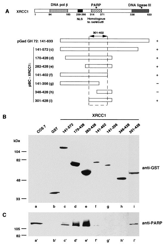
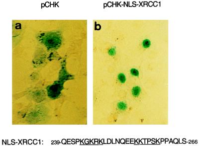
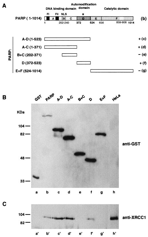
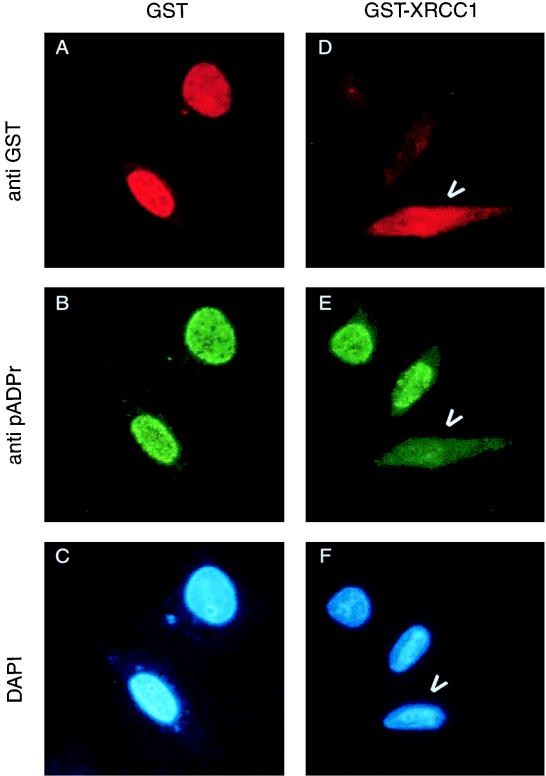
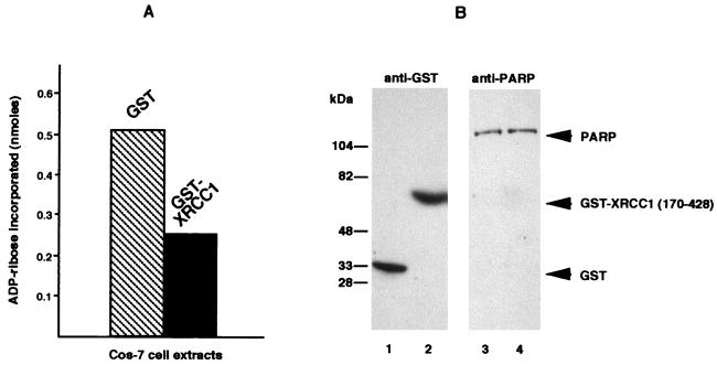
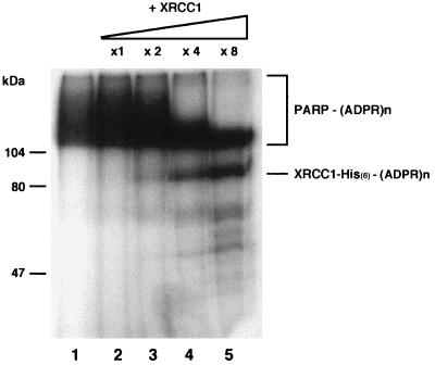
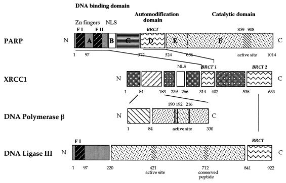
Poly(ADP-ribose) polymerase (PARP; EC 2.4.2.30) is a zinc-finger DNA-binding protein that detects and signals DNA strand breaks generated directly or indirectly by genotoxic agents. In response to these breaks, the immediate poly(ADP-ribosyl)ation of nuclear proteins involved in chromatin architecture and DNA metabolism converts DNA damage into intracellular signals that can activate DNA repair programs or cell death options. To have greater insight into the physiological function of this enzyme, we have used the two-hybrid system to find genes encoding proteins putatively interacting with PARP. We have identified a physical association between PARP and the base excision repair (BER) protein XRCC1 (X-ray repair cross-complementing 1) in the Saccharomyces cerevisiae system, which was further confirmed to exist in mammalian cells. XRCC1 interacts with PARP by its central region (amino acids 301 to 402), which contains a BRCT (BRCA1 C terminus) module, a widespread motif in DNA repair and DNA damage-responsive cell cycle checkpoint proteins. Overexpression of XRCC1 in Cos-7 or HeLa cells dramatically decreases PARP activity in vivo, reinforcing the potential protective function of PARP at DNA breaks. Given that XRCC1 is also associated with DNA ligase III via a second BRCT module and with DNA polymerase beta, our results provide strong evidence that PARP is a member of a BER multiprotein complex involved in the detection of DNA interruptions and possibly in the recruitment of XRCC1 and its partners for efficient processing of these breaks in a coordinated manner. The modular organizations of these interactors, associated with small conserved domains, may contribute to increasing the efficiency of the overall pathway.


Figures

FIG. 1
FIG. 1
Interaction of PARP with GST-tagged XRCC1 domains. (A) Modular organization of XRCC1. Amino acids 84 to 183 make up the region that interacts with DNA polymerase (pol) β (25); amino acids 538 to 633 make up the region that interacts with DNA ligase III (37); amino acids 301 to 402 make up the region that interacts with PARP (cf. panels B and C); and amino acids 239 to 266 make up the NLS (cf. Fig. 2). The asterisks indicate the two BRCT modules (4, 9). The portion of XRCC1 encoded by the cDNA clone isolated in the two-hybrid procedure and the GST-tagged XRCC1 deletion mutants expressed in Cos-7 cells are also diagrammed. Expressed GST-fusion proteins and interacting endogenous proteins (PARP) were selectively extracted and analyzed by Western blotting as described in Materials and Methods with successively anti-GST (B) and anti-PARP (C) antibodies. Lanes a and a′ contain the total extract of control untransfected Cos-7 cells.
FIG. 2
FIG. 2
XRCC1 NLS. Cos-7 cells were transfected with the empty vector pCHK (a) or with the construct pCHK-NLS-XRCC1-(239–266), which encodes the XRCC1 bipartite NLS fused to β-galactosidase (b). The subcellular localization of the recombinant proteins was assessed by histochemical staining with X-Gal (5-bromo-4-chloro-3-indolyl-β-d-galactopyranoside). Micrographs of Cos-7 cells transfected with the XRCC1 putative NLS exclusively displayed blue nuclei (b).
FIG. 3
FIG. 3
Interaction of XRCC1 with GST-PARP and PARP functional domains. (A) Modular organization of PARP (diagram b) and truncated forms of PARP (diagrams c to g) expressed as GST-fusion proteins in HeLa cells. The asterisk indicates the BRCT motif present in domain D of PARP; FI and FII correspond to the PARP zinc fingers (4, 9). Fusion proteins and interacting endogenous proteins (XRCC1) were selectively extracted and analyzed by Western blotting as described in Materials and Methods with successively anti-GST (B) and anti-XRCC1 (C) antibodies. Lanes h and h′ contain total extract of control untransfected HeLa cells.
FIG. 4
FIG. 4
In vivo negative regulation of PARP activity by XRCC1. HeLa cells expressing either GST alone (A to C) or GST-XRCC1 (D to F) were treated with H2O2 for 10 min. After methanol-acetone fixation, the cells were incubated with a mixture of monoclonal antibodies against GST and against poly(ADP-ribose) (pADPr). The primary antibodies were detected with Texas red or fluorescein isothiocyanate-conjugated secondary antibodies. Arrowheads point out cells expressing GST-XRCC1 and lacking PARP activity (D to F). DAPI, 4′,6-diamidino-2-phenylindole.
FIG. 5
FIG. 5
In vitro negative regulation of PARP activity by XRCC1. (A) PARP activities were determined in total extracts of Cos-7 cells expressing either GST or GST–XRCC1-(170–428), as described in Materials and Methods. (B) Immunoblot detection of GST (lanes 1 and 2) and PARP (lanes 3 and 4) in extracts from Cos-7 cells transfected either with pBC-NLS (lanes 1 and 3) or with pBC XRCC1-(170–428) (lanes 2 and 4).
FIG. 6
FIG. 6
ADP-ribosylation of XRCC1 and its effect on PARP auto-poly(ADP-ribosylation). Incubations of XRCC1 with 300 ng of purified PARP were performed with 1 μM [32P]NAD for 2 min at 25°C under standard conditions (38). Acid-insoluble products were separated by gel electrophoresis and revealed by autoradiography of the stained and dried gel. Lane 1, PARP alone; lanes 2 to 5, purified His-tagged XRCC1 added at the indicated molar ratios with respect to PARP. (ADPR)n, ADP-ribosylation.
FIG. 7
FIG. 7
Network of interactions between enzymes and factors participating in the BER pathway. BRCT modules involved in PARP-XRCC1 and in DNA ligase III-XRCC1 (37) interactions are indicated. XRCC1 contacts DNA polymerase β (25) by its N-terminal region. PARP and DNA ligase III have the same nick detection motif (zinc finger F1 [F I], amino acids 1 to 97).

References

    1. Althaus F R, Richter C. ADP-ribosylation of proteins. Enzymology and biological significance. Mol Biol Biochem Biophys. 1987;37:1–237. - PubMed
    1. Barlow C, Hirotsune S, Paylor R, Liyanage M, Eckhaus M, Collins F, Shilo Y, Crawley J N, Ried T, Tagle D, Wynshaw-Boris A. Atm-deficient mice: a paradigm of ataxia telangectasia. Cell. 1996;86:159–171. - PubMed
    1. Biedermann K A, Sun J, Giaccia A J, Tosto L M, Brown J M. Scid mutation in mice confers hypersensitivity to ionizing radiation and a deficiency in DNA double-strand break repair. Proc Natl Acad Sci USA. 1991;88:1394–1397. - PMC - PubMed
    1. Bork P, Hofmann K, Bucher P, Neuwald A F, Altschul S F, Koonin E V. A superfamily of conserved domains in DNA damage-responsive cell cycle checkpoint proteins. FASEB J. 1997;11:68–76. - PubMed
    1. Brookman K W, Tebbs R S, Allen S A, Tucker J D, Swiger R R, Lamerdin J E, Carrano A V, Thompson L H. Isolation and characterization of mouse Xrcc-1, a DNA repair gene affecting ligation. Genomics. 1994;22:180–188. - PubMed

Publication types