. 1998 Jun;18(6):3586-95.
doi: 10.1128/MCB.18.6.3586.

Characterization of interactions between the mammalian polycomb-group proteins Enx1/EZH2 and EED suggests the existence of different mammalian polycomb-group protein complexes

Affiliations

Characterization of interactions between the mammalian polycomb-group proteins Enx1/EZH2 and EED suggests the existence of different mammalian polycomb-group protein complexes

R G Sewalt et al. Mol Cell Biol. 1998 Jun.

Abstract

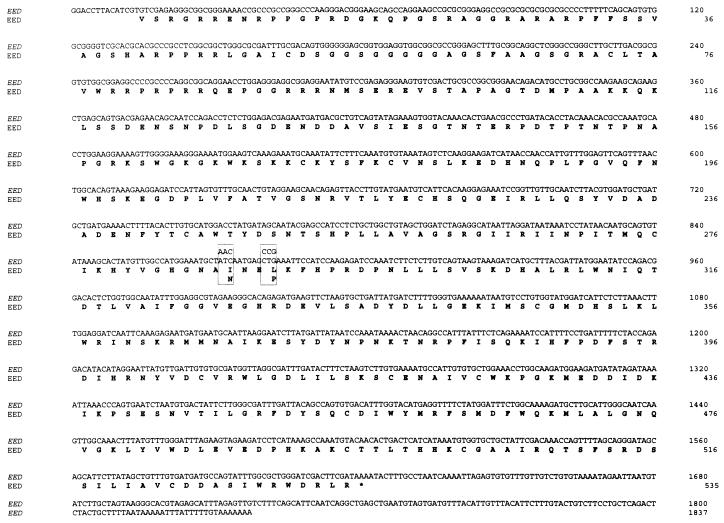
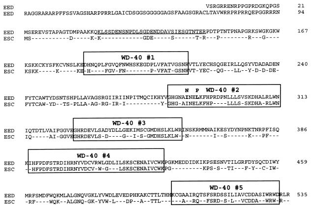
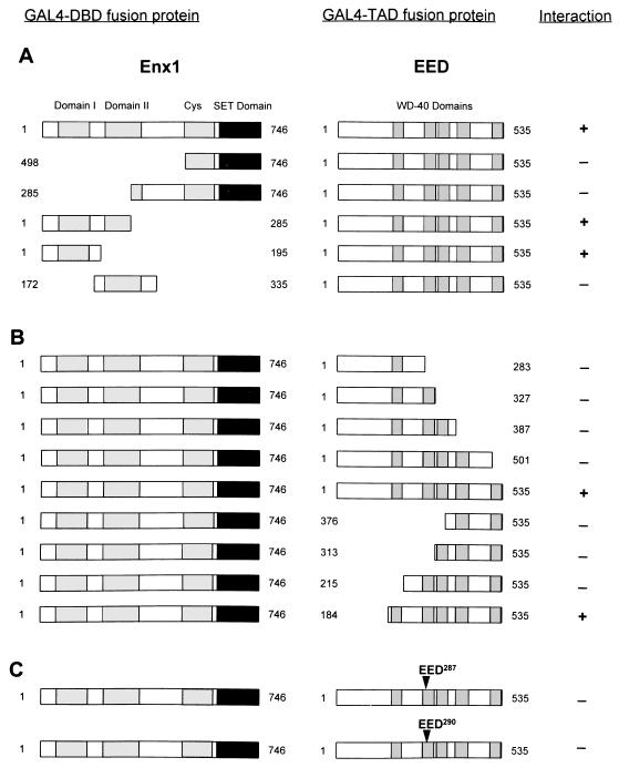
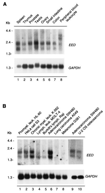
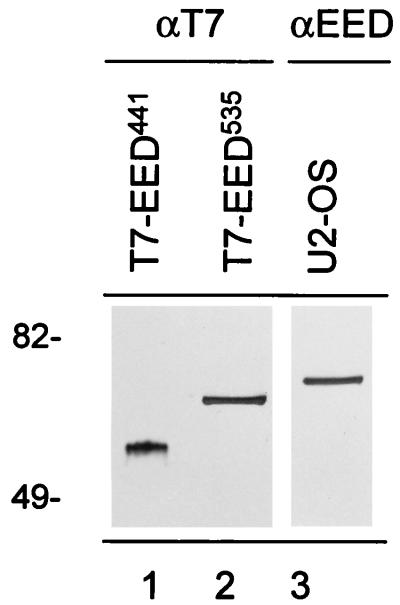
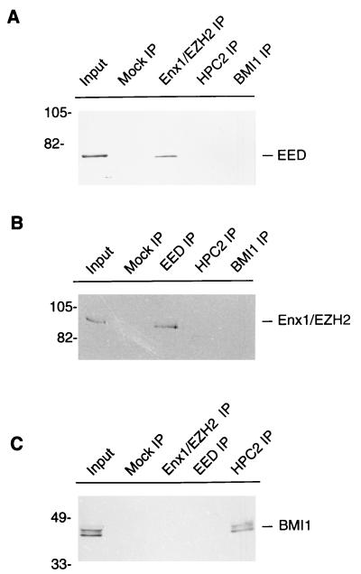
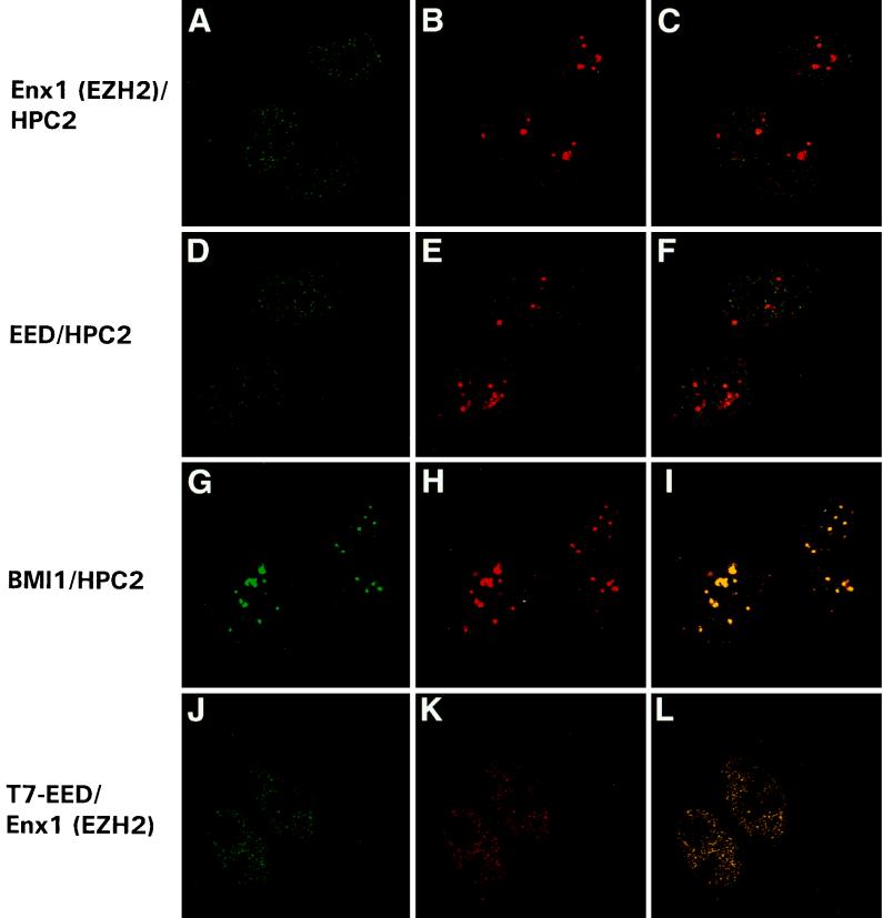
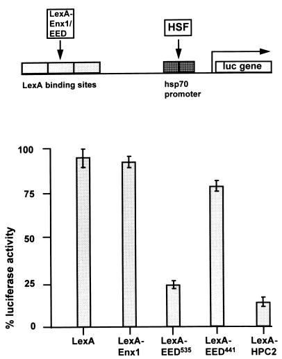
In Drosophila melanogaster, the Polycomb-group (PcG) and trithorax-group (trxG) genes have been identified as repressors and activators, respectively, of gene expression. Both groups of genes are required for the stable transmission of gene expression patterns to progeny cells throughout development. Several lines of evidence suggest a functional interaction between the PcG and trxG proteins. For example, genetic evidence indicates that the enhancer of zeste [E(z)] gene can be considered both a PcG and a trxG gene. To better understand the molecular interactions in which the E(z) protein is involved, we performed a two-hybrid screen with Enx1/EZH2, a mammalian homolog of E(z), as the target. We report the identification of the human EED protein, which interacts with Enx1/EZH2. EED is the human homolog of eed, a murine PcG gene which has extensive homology with the Drosophila PcG gene extra sex combs (esc). Enx1/EZH2 and EED coimmunoprecipitate, indicating that they also interact in vivo. However, Enx1/EZH2 and EED do not coimmunoprecipitate with other human PcG proteins, such as HPC2 and BMI1. Furthermore, unlike HPC2 and BMI1, which colocalize in nuclear domains of U-2 OS osteosarcoma cells, Enx1/EZH2 and EED do not colocalize with HPC2 or BMI1. Our findings indicate that Enx1/EZH2 and EED are members of a class of PcG proteins that is distinct from previously described human PcG proteins.


Figures

FIG. 1
FIG. 1
Nucleotide sequence of EED and its predicted amino acid sequence. The point mutations (bp 872 and 881) in eed that are found in the mutant eed mice are boxed. The stop codon of the EED gene is indicated with an asterisk.
FIG. 2
FIG. 2
Comparison of the EED/eed protein with the Drosophila PcG protein esc. Identical amino acids are shown; nonidentical amino acids are indicated with a dash. The five WD-40 repeats are indicated with boxes. A putative PEST sequence is underlined. The point mutations (aa 287 and 290) in eed that are found in the mutant eed mice are shaded in the boxed WD-40 domain 2.
FIG. 3
FIG. 3
Mapping of interaction domains between Enx1 and EED. (A) Indicated portions of Enx1 were fused to the GAL4-DBD (GAL4-DBD fusion protein). These Enx1 regions include homology domains I (aa 94 to 159), homology domain II (aa 218 to 329), a cysteine-rich domain (aa 498 to 612), and the SET domain (aa 613 to 742). Constructs that encompass different portions of the Enx1 protein are indicated. The plasmids were cotransformed with full-length EED (aa 1 to 535), which is fused to the GAL4-TAD (GAL4-TAD fusion protein). Interactions were positive (+) when the transformants were able to grow on selective medium lacking histidine and when they were also β-galactose positive. (B) Full-length Enx1 (aa 1 to 746) fused to the GAL4-DBD was tested for interaction against indicated portions of EED fused to the GAL4-TAD. (C) Indicated point mutations in the second WD-40 domain of EED were made and tested against the full-length Enx1 protein.
FIG. 4
FIG. 4
Expression patterns of EED in human tissues (A) and in human cancer cell lines (B). (A) Expression levels in spleen (lane 1), thymus (lane 2), prostate (lane 3), testis (lane 4), ovary (lane 5), small intestine (lane 6), colon (lane 7), and peripheral blood leukocytes (lane 8). (B) Expression levels in promyelocytic leukemia HL-60 (lane 1), HeLa cell S3 (lane 2), chronic myelogenous leukemia K-562 (lane 3), lymphoblastic leukemia MOLT-4 (lane 4), Burkitt’s lymphoma Raji (lane 5), colorectal adenocarcinoma SW480 (lane 6), lung carcinoma A549 (lane 7), and melanoma G361 (lane 8) cell lines. Lanes 1 to 8 represent a commercially obtained Northern blot. We also isolated and blotted poly(A)+ RNA from U-2 OS (lane 10). To allow comparison with the commercial multiple-tissue Northern blot, we isolated and blotted poly(A)+ RNA from SW480 cells (lane 9). The filters were rehybridized with a probe for glyceraldehyde-3-phosphate dehydrogenase (GAPDH) to verify the loading of RNA in each lane.
FIG. 5
FIG. 5
Comparison between different EED proteins. Two T7-tagged EED constructs which potentially encode the EED441 protein and EED535 protein were made. U-2 OS cells were transfected with T7-EED441 (lane 1) and T7-EED535 (lane 2) cDNAs, and the respective cell lysates were analyzed by Western blotting. Blots were probed with a mouse monoclonal antibody against T7 (αT7; lanes 1 and 2). The endogenous EED protein was detected in a cell lysate of U-2 OS cells (lane 3), using an affinity-purified antibody against EED (αEED). Molecular weights are indicated in thousands.
FIG. 6
FIG. 6
Enx1/EZH2 and EED coimmunoprecipitate from extracts of U-2 OS cells. IP experiments were performed with extracts of U-2 OS human osteosarcoma cells. (A) IP performed with polyclonal rabbit antibodies against Enx1/EZH2 (Enx1/EZH2 IP), HPC2 (HPC2 IP), and BMI1 (BMI1 IP) or with preimmune serum (Mock IP). The resulting IPs were Western blotted and incubated with rabbit anti-EED antibody. The 68-kDa EED protein was detected in the U-2 OS cell extract (Input) and in the Enx1/EZH2 IP but not in the HPC2 IP and BMI1 IP. (B) IP performed with polyclonal rabbit antibodies against EED (EED IP), HPC2 (HPC2 IP), and BMI1 (BMI1 IP) or with preimmune serum (Mock IP). The resulting IPs were Western blotted and incubated with rabbit anti-Enx1 antibody. The approximately 90-kDa Enx1/EZH2 protein was detected in the U-2 OS cell extract (Input) and in the EED IP, but not in the HPC2 IP and BMI1 IP. (C) IP performed using polyclonal rabbit antibodies against Enx1/EZH2 (Enx1/EZH2 IP), EED (EED IP), and HPC2 (HPC2 IP) or with preimmune serum (Mock IP). The resulting IPs were Western blotted and incubated with chicken anti-BMI1 antibody. The approximately 44- to 47-kDa BMI1 protein was detected in the U-2 OS cell extract (Input) and in the HPC2 IP, but not in the Enx1/EZH2 IP and the EED IP. Molecular weights are indicated in thousands.
FIG. 7
FIG. 7
Enx1/EZH2 and EED do not colocalize with the PcG protein HPC2 in nuclear domains of U-2 OS cells. Confocal single optical sections of double-labeled cells are presented. (A to C) Rabbit anti-Enx1/EZH2 and chicken anti-HPC2 double labeling. Enx1/EZH2 (A), like HPC2 (B), is homogeneously distributed in the nucleus, but unlike HPC2, Enx1/EZH2 is not concentrated in large, brightly labeled domains (B and C). (D to F) Rabbit anti-EED and chicken anti-HPC2 double labeling. EED (D), like HPC2 (E), is homogeneously distributed in the nucleus, but unlike HPC2, EED is not concentrated in large, brightly labeled domains (E and F). Rabbit anti-BMI1 (G) and chicken anti-HPC2 (H) double labeling demonstrates colocalization of HPC2 and BMI1 in the large bright domains (I) (indicated by yellow). We transiently transfected U-2 OS cells with the T7-tagged EED535 protein. Double labeling was performed with a mouse monoclonal antibody against T7 (J) and the affinity-purified rabbit antibody against Enx1/EZH2 (K). We observed complete colocalization between T7-EED535 and Enx1/EZH2 (L).
FIG. 8
FIG. 8
Repression of HSF-induced LUC gene activity by Enx1 and EED. Activation of LUC reporter expression is maximally induced by endogenous HSF in the absence of any LexA fusion protein and was set at 100%. LUC activities in cells cotransfected with other plasmids were expressed as a percentage of this control value. Bars represent the average degree of repression by LexA, LexA-Enx1, LexA-EED535, LexA-EED441, or LexA-HPC2 in three independent experiments (mean ± standard error of the mean).

References

    1. Alkema M J, Wiegant J, Raap A K, Berns A, van Lohuizen M. Characterization and chromosomal localization of the human proto-oncogene BMI-1. Hum Mol Genet. 1993;2:1597–1603. - PubMed
    1. Alkema M J, Bronk M, Verhoeven E, Otte A P, van’t Veer L J, Berns A, van Lohuizen M. Identification of Bmi1-interacting proteins as constituents of a multimeric mammalian Polycomb complex. Genes Dev. 1997;11:226–240. - PubMed
    1. Bunker C A, Kingston R E. Transcriptional repression by Drosophila and mammalian Polycomb group proteins in transfected mammalian cells. Mol Cell Biol. 1994;14:1721–1732. - PMC - PubMed
    1. Carrington E A, Jones R S. The Drosophila Enhancer of zeste gene encodes a chromosomal protein: examination of wild-type and mutant protein distribution. Development. 1996;122:4073–4083. - PubMed
    1. Chang Y-L, King B O, O’Connor M, Mazo A, Huang D-H. Functional reconstruction of trans regulation of the Ultrabithorax promoter by the products of two antagonistic genes, trithorax and Polycomb. Mol Cell Biol. 1995;15:6601–6612. - PMC - PubMed

MeSH terms

Associated data