. 1998 May 18;141(4):929-42.
doi: 10.1083/jcb.141.4.929.

Lipid domain structure of the plasma membrane revealed by patching of membrane components

Affiliations

Lipid domain structure of the plasma membrane revealed by patching of membrane components

T Harder et al. J Cell Biol. .

Abstract

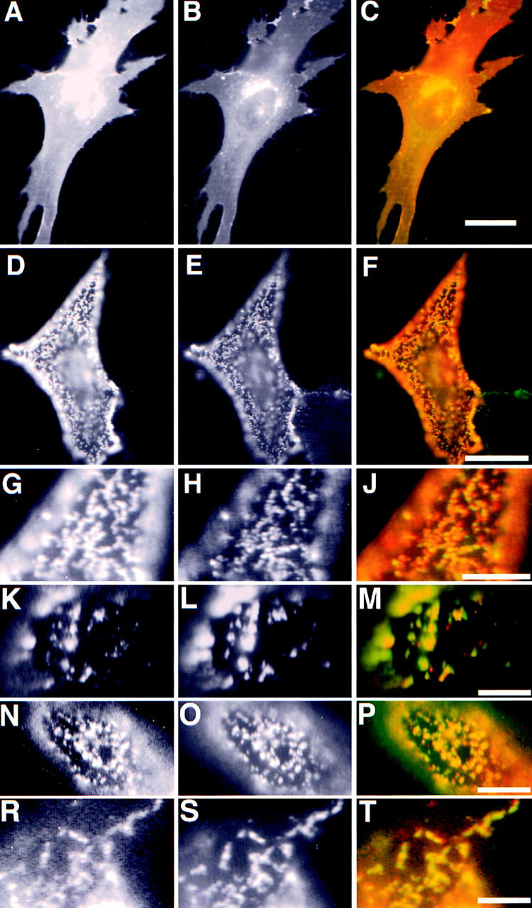
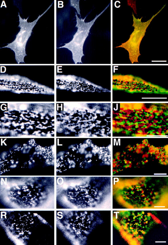
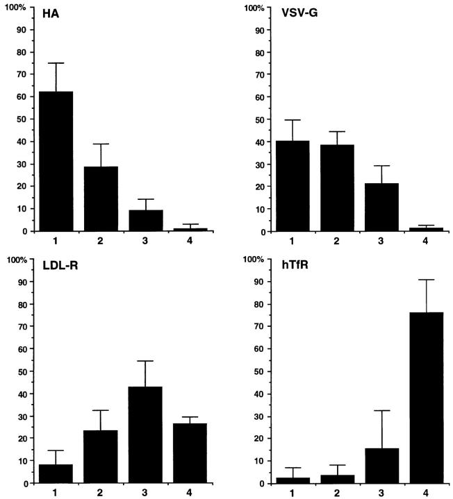
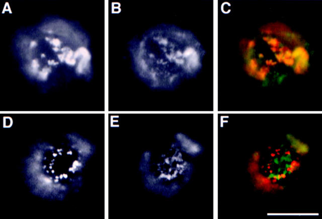
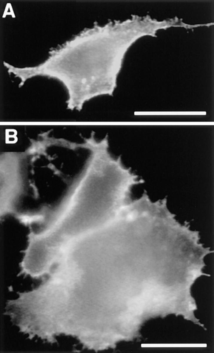
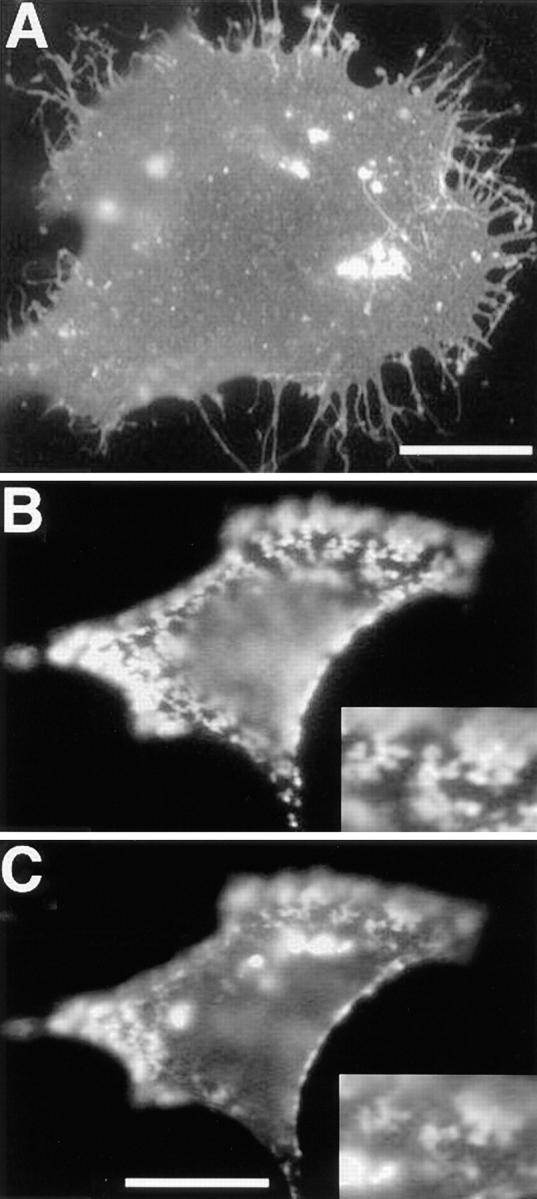
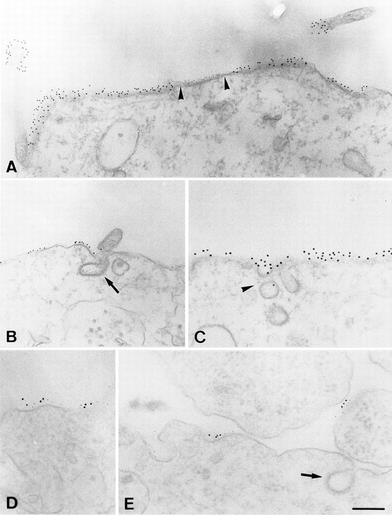
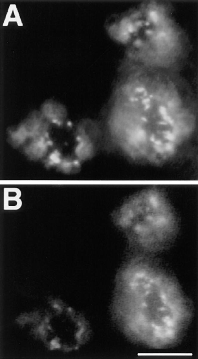
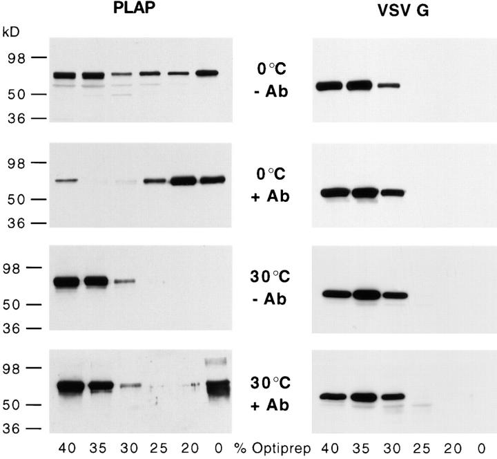
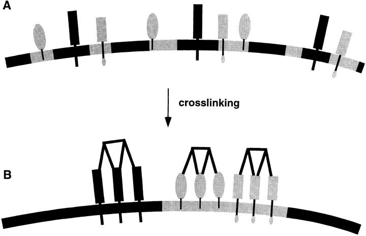
Lateral assemblies of glycolipids and cholesterol, "rafts," have been implicated to play a role in cellular processes like membrane sorting, signal transduction, and cell adhesion. We studied the structure of raft domains in the plasma membrane of non-polarized cells. Overexpressed plasma membrane markers were evenly distributed in the plasma membrane. We compared the patching behavior of pairs of raft markers (defined by insolubility in Triton X-100) with pairs of raft/non-raft markers. For this purpose we cross-linked glycosyl-phosphatidylinositol (GPI)-anchored proteins placental alkaline phosphatase (PLAP), Thy-1, influenza virus hemagglutinin (HA), and the raft lipid ganglioside GM1 using antibodies and/or cholera toxin. The patches of these raft markers overlapped extensively in BHK cells as well as in Jurkat T-lymphoma cells. Importantly, patches of GPI-anchored PLAP accumulated src-like protein tyrosine kinase fyn, which is thought to be anchored in the cytoplasmic leaflet of raft domains. In contrast patched raft components and patches of transferrin receptor as a non-raft marker were sharply separated. Taken together, our data strongly suggest that coalescence of cross-linked raft elements is mediated by their common lipid environments, whereas separation of raft and non-raft patches is caused by the immiscibility of different lipid phases. This view is supported by the finding that cholesterol depletion abrogated segregation. Our results are consistent with the view that raft domains in the plasma membrane of non-polarized cells are normally small and highly dispersed but that raft size can be modulated by oligomerization of raft components.


Figures

Figure 1
Figure 1
Copatching of cross-linked PLAP with influenza HA. The left column shows PLAP distribution in different experiments, the middle column HA distribution, and the right column the merge of the two signals. (A–C) Immunofluorescence of PLAP and wild-type HA on fixed BHK cells. (D–M) Copatching of PLAP with HA wild type. G–J show a detail of D–F. K–M show the copatching of PLAP and HA at 37°C. (N–P) Copatching of PLAP with HA tail minus, and (R– T) HA 543Y. PLAP antibodies were detected using rhodamine anti–mouse (red), HA antibodies using FITC anti–rabbit-labeled (green) secondary antibodies. Bars: (C) 10 μm; (F) 5 μm; (J, M, P, and T) 3 μm.
Figure 2
Figure 2
Patching of PLAP and transmembrane proteins hTfR, hTfR del 5–41, LDL-R YA18, and VSV-G. The left column shows PLAP distribution. The middle column shows distribution of hTfR (B, E, H, and L), LDL-R YA 18 mutant (O), and VSV-G (S). The right column shows the merge of the two signals. A–C shows immunofluorescence of PLAP (green) and hTfR (red) on fixed BHK cells. D–J shows segregation of patched PLAP (green) and hTfR (red). G–J shows a detail of D–F. K–M shows segregation patches of cross-linked PLAP (green) and hTfR del 5–41 (red) at 37°C. N–P shows patches of cross-linked PLAP (green) and LDL-R YA18 (red). R–T show patches of PLAP (red) and VSV-G (green). Bars: (C) 10 μm; (F) 5 μm; (J, M, P, and T) 3 μm.
Figure 3
Figure 3
Quantitation of copatching of influenza HA, VSV-G, LDL-R, and hTfR with cross-linked PLAP. Patches of the different membrane markers were scored into four categories: (1) copatching (>80% overlap); (2) partial copatching (clearly overlapping regions); (3) random distribution; (4) segregation. The percentages of cells falling into each category are expressed as averages and SD from at least four experiments.
Figure 4
Figure 4
Patching of GM1/cholera toxin with PLAP and hTfR del 5–41 in Jurkat T–lymphoma cells. FITC-CTx conjugate was used to patch and stain GM 1 (green). Respective mAbs against PLAP or hTfR were used followed by incubation with rhodamine-coupled secondary antibodies (red). PLAP (A) and GM1 (B) show extensive copatching (merge in C). Patched hTfR del 5–41 (D) and GM1 patches (E) are largely exclusive (merge in F). Bar, 5 μm.
Figure 5
Figure 5
PLAP and hTfR patching depends on membrane cholesterol. BHK cells transiently expressing PLAP (A) and hTfR (B) were depleted of membrane cholesterol using cyclodextrin extraction. PLAP and hTfR were cross-linked using anti-PLAP and anti-hTfR mAb, respectively, followed by rhodamine-coupled secondary antibodies. Patching of PLAP as well as hTfR is inhibited under these conditions. Bar, 5 μm.
Figure 6
Figure 6
Accumulation of fyn in membrane domains formed by patched PLAP. A shows distribution of overexpressed fyn in FA/ MeOH-fixed BHK cells. Patches of PLAP are shown in B, and the corresponding distribution of fyn in C. The inserts show a detail of the overlap between PLAP and fyn staining. The faint diffuse signal in the rhodamine (PLAP) channel that coincides with the intensely bright fyn aggregates (FITC) is most probably because of a small overflow from the FITC to the rhodamine channel. The overlap between the patched PLAP and the signal from the fyn immunofluorescence at the cell surface is however clearly independent from the intracellular fyn aggregates. Bars: (A) 10 μm; (C) 5 μm.
Figure 7
Figure 7
Electron microscopical analysis of patched membrane components. A and B show separation of PLAP patches (12-nm gold) and hTfR patches (6-nm gold). Arrowheads, the sharp boundaries between the patches. Arrows in B show a clathrin-coated invagination labeled for hTfR. C shows patches of PLAP surrounding caveolar-like, noncoated invaginations (arrowhead). D and E show copatching of HA (12-nm gold) and PLAP (6-nm gold) on smooth membrane regions. Arrow in E marks clathrin-coated invagination. Bars: (A–C) 250 nm; (D and E) 150 nM.
Figure 8
Figure 8
Copatching in T hybridoma cells. GM1 and Thy-1 copatching was induced in 2B2318 T–hybridoma cells by simultaneous incubation with anti–Thy-1 mAb and FITC-CTx followed by incubation with rhodamine anti–mouse antibodies. A depicts distribution Thy-1 patches and B distribution of the patches of GM1 CTx. Bar, 5 μm.
Figure 9
Figure 9
Stabilization of membrane domains by antibody cross-linking. BHK cells expressed PLAP after transient transfection or VSV-G introduced by VSV infection. PLAP (left) and VSV-G (right) were patched using an mAb against PLAP and polyclonal antibodies against VSV-G, respectively, followed by the respective secondary antibodies. Cells were subsequently lysed in 2% Triton X-100 at 4° or 30°C. Fractions of an Optiprep™–sucrose flotation step gradient were analyzed by Western blot using anti-PLAP and anti–VSV-G antibodies. Antibody-induced patching (+Ab) significantly increases the amount of PLAP associated to a Triton X-100–insoluble membrane fraction that floats to the interphase of 20% Optiprep™/10% sucrose and 0% Optiprep™/ 10% sucrose, whereas VSV-G remains in high density fraction irrespective whether it was cross-linked or not.
Figure 10
Figure 10
Bulk separation of membrane phases caused by clustering of membrane components. (A) Microdomains and membrane proteins in these domains are dispersed in the plasma membrane. (B) Cross-linking generates large and stabilized membrane domains that coalesce to form patches. If two membrane components share a preference for a lipid environment such as raft microdomains the markers will copatch into tightly associated domains. If two markers partition into different membrane environments such as raft and non-raft markers the patches will be separated.

Similar articles

Cited by

References

    1. Ahmed SN, Brown DA, London E. On the origin of sphingolipid/ cholesterol-rich detergent-insoluble cell membranes: physiological concentrations of cholesterol and sphingolipid induce formation of a detergent insoluble, liquid ordered lipid phase in model membranes. Biochemistry. 1997;36:10944–10953. - PubMed
    1. Blackman MA, Lund FE, Surman S, Corley RB, Woodland DL. Major histocompatibility complex restricted recognition of retroviral superantigens by V β 17+ T cells. J Exp Med. 1992;176:275–280. - PMC - PubMed
    1. Brewer CB, Roth MG. A single amino acid change in the cytoplasmic domain alters the polarized delivery of influenza virus hemagglutinin. J Cell Biol. 1991;114:413–421. - PMC - PubMed
    1. Brown DA. Interactions between GPI-anchored proteins and membrane lipids. Trends Cell Biol. 1992;2:338–343. - PubMed
    1. Brown DA. The tyrosine kinase connection: how GPI-anchored proteins activate T cells. Curr Opin Immunol. 1993;5:349–354. - PubMed

Publication types

MeSH terms