. 1998 May 18;141(4):967-77.
doi: 10.1083/jcb.141.4.967.

Analysis of the Saccharomyces spindle pole by matrix-assisted laser desorption/ionization (MALDI) mass spectrometry

Affiliations

Analysis of the Saccharomyces spindle pole by matrix-assisted laser desorption/ionization (MALDI) mass spectrometry

P A Wigge et al. J Cell Biol. .

Abstract

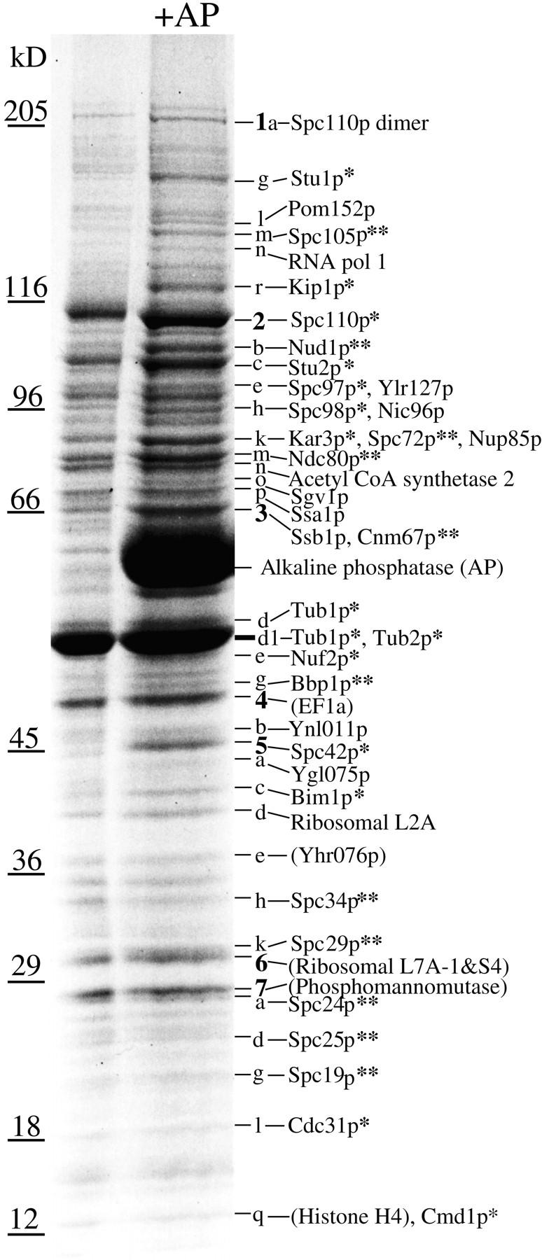
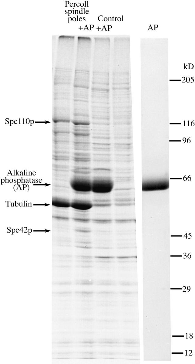
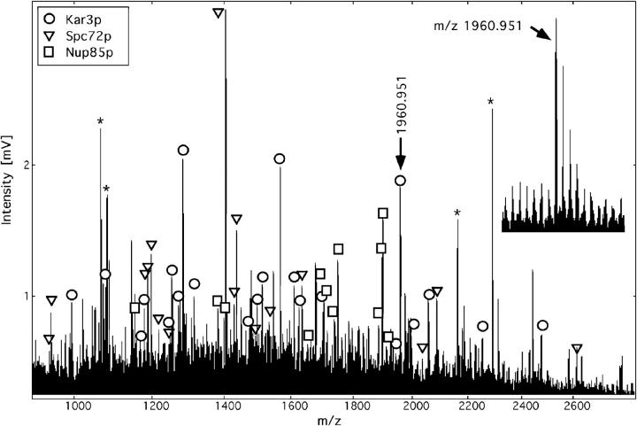
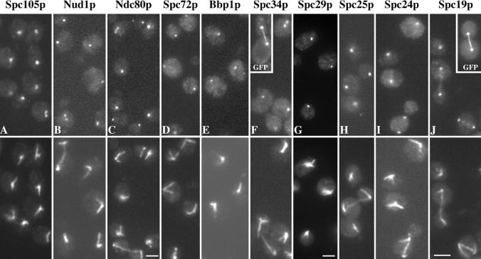
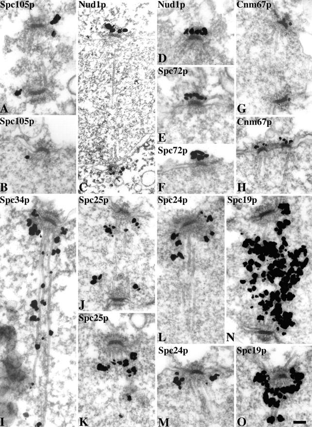
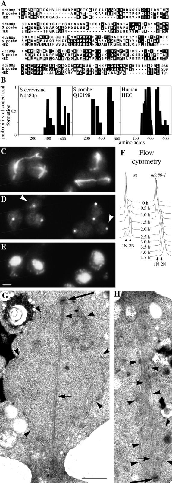
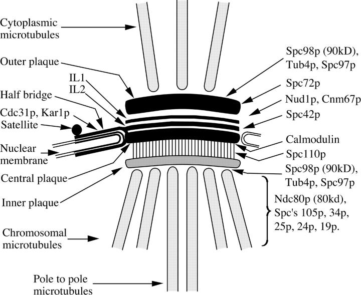
A highly enriched spindle pole preparation was prepared from budding yeast and fractionated by SDS gel electrophoresis. Forty-five of the gel bands that appeared enriched in this fraction were analyzed by high-mass accuracy matrix-assisted laser desorption/ ionization (MALDI) peptide mass mapping combined with sequence database searching. This identified twelve of the known spindle pole components and an additional eleven gene products that had not previously been localized to the spindle pole. Immunoelectron microscopy localized eight of these components to different parts of the spindle. One of the gene products, Ndc80p, shows homology to human HEC protein (Chen, Y., D.J. Riley, P-L. Chen, and W-H. Lee. 1997. Mol. Cell Biol. 17:6049-6056) and temperature-sensitive mutants show defects in chromosome segregation. This is the first report of the identification of the components of a large cellular organelle by MALDI peptide mapping alone.


Figures

Figure 3
Figure 3
Identification of proteins in Coomassie-stained SDS gel bands from AP-treated yeast spindle poles by MALDI mass mapping. Single asterisk, a spindle pole component already identified; double asterisk, a new spindle pole component. Bands were labeled by giving all the prominent bands numbers (boldface) then bands underneath were given that number and a letter in order of decreasing size until the next prominent numbered band. Identifications in parentheses are from prominent bands that are not enriched in the spindle pole preparation. The faint Coomassie-stained bands between 205 and 116 kD contain ∼10–20 ng of protein by comparison with BSA standards.
Figure 1
Figure 1
SDS gel of two adjacent fractions from the final Percoll gradient. The first two lanes contain most of the spindle poles treated with and without alkaline phosphatase (AP), the next two lanes labeled control contain the next gradient fraction which has less than 15% of the poles present in the first fraction. A large number of gel bands are specifically enriched in the spindle pole fraction. The last lane is AP alone.
Figure 2
Figure 2
Identification of the proteins Kar3p, Spc72p, and Nup85p in band 2k by MALDI peptide mass mapping and iterative database searching using a peptide mass error of less than 30 ppm. Asterisk, matrix or trypsin autolysis peaks used as internal calibration peaks; inset, data quality obtained for the ion signal at m/z 1960, mass resolution of 10,000 (full width at half maximum) allowed monoisotopic mass determination with a mass error of only 11 ppm. The measured masses (monoisotopic protonated peptide molecular weight) of the three proteins identified together with the deviations from the matched mass and any amino acid modifications in brackets were as follows: Kar3p: 992.627 (0.027), 1077.628 (−0.002), 1170.580 (0.034), 1178.627 (0.010), 1248.654 (0.011), 1252.675 (0.011), 1272.706 (−0.010), 1284.643 (−0.005, methionine sulphoxide), 1314.669 (−0.011), 1475.793 (−0.013), 1498.724 (0.014, methionine sulphoxide), 1513.832 (0.021), 1568.788 (0.029), 1610.862 (−0.003), 1626.839 (0.033, methionine sulphoxide), 1704.891 (0.021), 1945.031 (−0.004), 1960.951 (−0.021), 1997.982 (−0.014), 2057.999 (0.011), 2253.089 (−0.033), 2476.233 (−0.017, methionine sulphoxide); Yal047p/ Spc72p: 939.571 (0.009), 943.592 (−0.001), 1187.635 (−0.003), 1188.617 (0.032), 1196.685 (0.018), 1218.662 (−0.025), 1246.659 (−0.020), 1406.755 (0.028), 1429.754 (−0.010), 1437.828 (−0.018), 1486.812 (0.001), 1534.814 (−0.008), 1635.867 (−0.022), 2036.030 (0.037), 2087.030 (0.004), 2613.342 (0.053); Nup85p: 1147.631 (−0.008), 1383.750 (−0.006), 1399.740 (0.018), 1653.830 (−0.037), 1680.841 (0.014), 1705.901 (−0.026), 1738.873 (0.012), 1751.876 (−0.020), 1885.920 (−0.016), 1898.983 (−0.018), 1899.954 (0.002, S-acrylamidocysteine), 1934.916 (−0.049). The particular peptides identified in each protein can be obtained by entering the measured masses into the PeptideSearch software available at http://www.mann.embl-heidelberg.de
Figure 4
Figure 4
Immunofluorescent staining of HA-tagged spindle pole components with mouse anti-HA mAb 12CA5 (top) and rabbit anti-tubulin (bottom). Insets, F and I, unfixed diploid GFP-labeled cells at half magnification. Bars, 2.5 μm, except C and G where bar is 2 μm.
Figure 5
Figure 5
ImmunoEM using preembedding labeling and Nanogold-silver intensification of eight of the spindle pole components. Spc105p was tagged with HA and all the other components with GFP. Bar, 0.1 μm.
Figure 6
Figure 6
Characterization of Ndc80p. (A) Homology between the NH2-terminal domains of Ndc80p, S. pombe Q10198, and human HEC; (B), comparison of the coiled-coil domains (determined by Paircoil) of the same three proteins; (C–E), immunofluorescent staining of ndc80-1 after 2 h at 36°C with anti-tubulin (C); anti-Tub4p to stain the SPBs (D); and DAPI to stain DNA (E). (D) White arrowheads, SPBs not associated with chromosomes; (F) flow cytometry to measure DNA replication in wild-type and ndc10-1 cells synchronized by elutriation and released at 36°C; (G and H), electron micrographs of serial thin sections from the same large budded ndc80-1 cell synchronized by α-factor and released at 36°C for 2.5 h. G shows one end of a postanaphase spindle and H the other end of the same spindle in the adjacent section. Large arrows, SPBs; small arrows, microtubules; arrowheads, nuclear pores and nuclear membrane. Bars: (C–E) 2 μm; (G and H) 0.5 μm.
Figure 7
Figure 7
Diagram of the localizations of spindle pole and SPB components described in this article. Other spindle pole components already localized by immunoEM are also shown. The localizations of the components Ndc80p and Spc's 105p, 34p, 25p, 24p, and 19p have not been established precisely relative to the nuclear side of the SPB. Ndc80p and Spc's 34p and 19p are probably spindle-associated since they can localize along the length of the spindle. IL1 and IL2, intermediate lines 1 and 2 (Bullitt et al., 1997). In thin sections IL2 is not visible since it appears fused to the central plaque.

References

    1. Aitchison JD, Rout MP, Marelli M, Blobel G, Wozniak RW. Two novel related yeast nucleoporins Nup170p and Nup155p: Complementation with the vertebrate homologue Nup155p and functional interactions with the yeast nuclear pore-membrane protein Pom152p. J Cell Biol. 1995;131:1133–1148. - PMC - PubMed
    1. Altschul SF, Gish W, Miller W, Myers EW, Lipman DJ. Basic local alignment search tool. J Mol Biol. 1990;215:403–410. - PubMed
    1. Baudin A, Ozier K, Denouel A, Lacroute F, Cullin C. A simple and efficient method for direct gene deletion in Saccharomyces cerevisiae. . Nucl Acids Res. 1993;21:3329–3330. - PMC - PubMed
    1. Beinhauer JD, Hagan IM, Hegemann JH, Fleig U. Mal3, the fission yeast homologue of the human APC-interacting protein EB-1 is required for microtubule integrity and the maintenance of cell form. J Cell Biol. 1997;139:717–728. - PMC - PubMed
    1. Bouckson-Castaing V, Moudjou M, Ferguson DJP, Mucklow S, Belkaid Y, Milon G, Crocker PR. Molecular characterization of ninein, a new coiled-coil protein of the centrosome. J Cell Sci. 1996;109:179–190. - PubMed

Publication types

MeSH terms