. 1998 May 15;12(10):1453-63.
doi: 10.1101/gad.12.10.1453.

Regulation of the fission yeast transcription factor Pap1 by oxidative stress: requirement for the nuclear export factor Crm1 (Exportin) and the stress-activated MAP kinase Sty1/Spc1

Affiliations

Regulation of the fission yeast transcription factor Pap1 by oxidative stress: requirement for the nuclear export factor Crm1 (Exportin) and the stress-activated MAP kinase Sty1/Spc1

W M Toone et al. Genes Dev. .

Erratum in

  • Genes Dev 1998 Aug 15;12(16):2650

Abstract

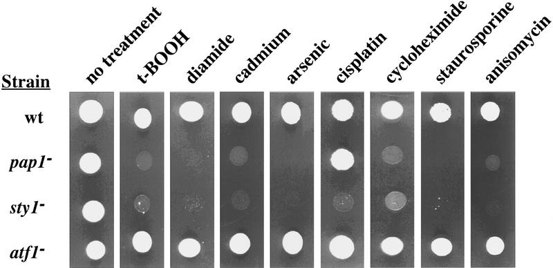
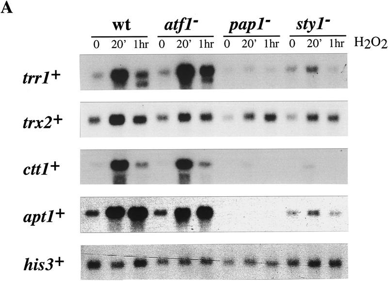
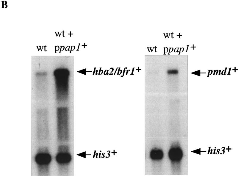
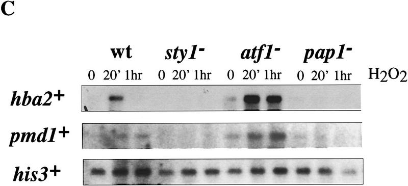
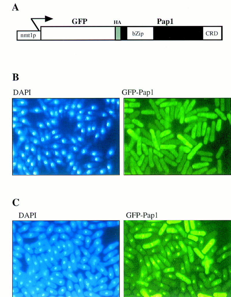
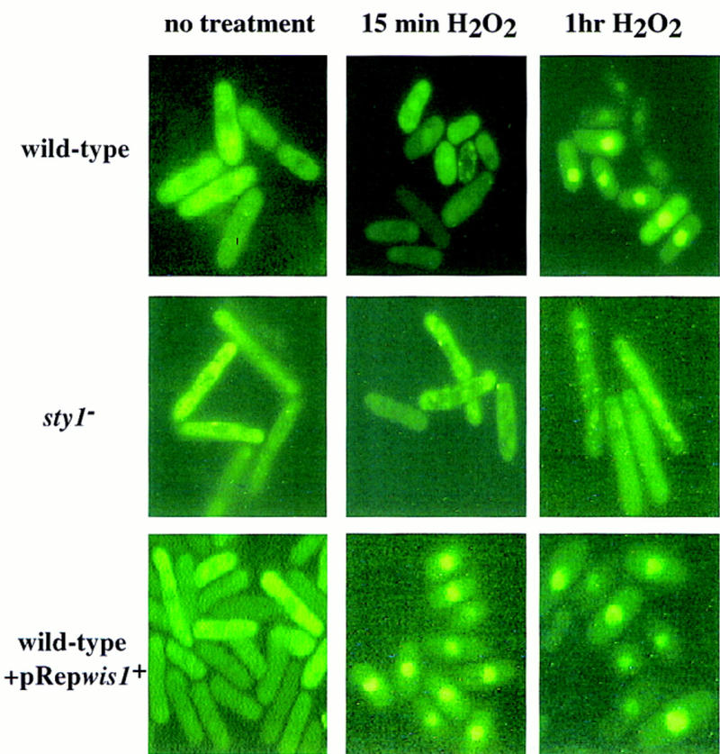
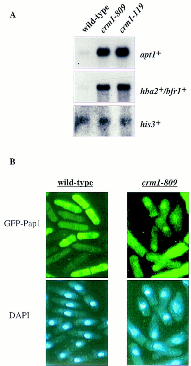
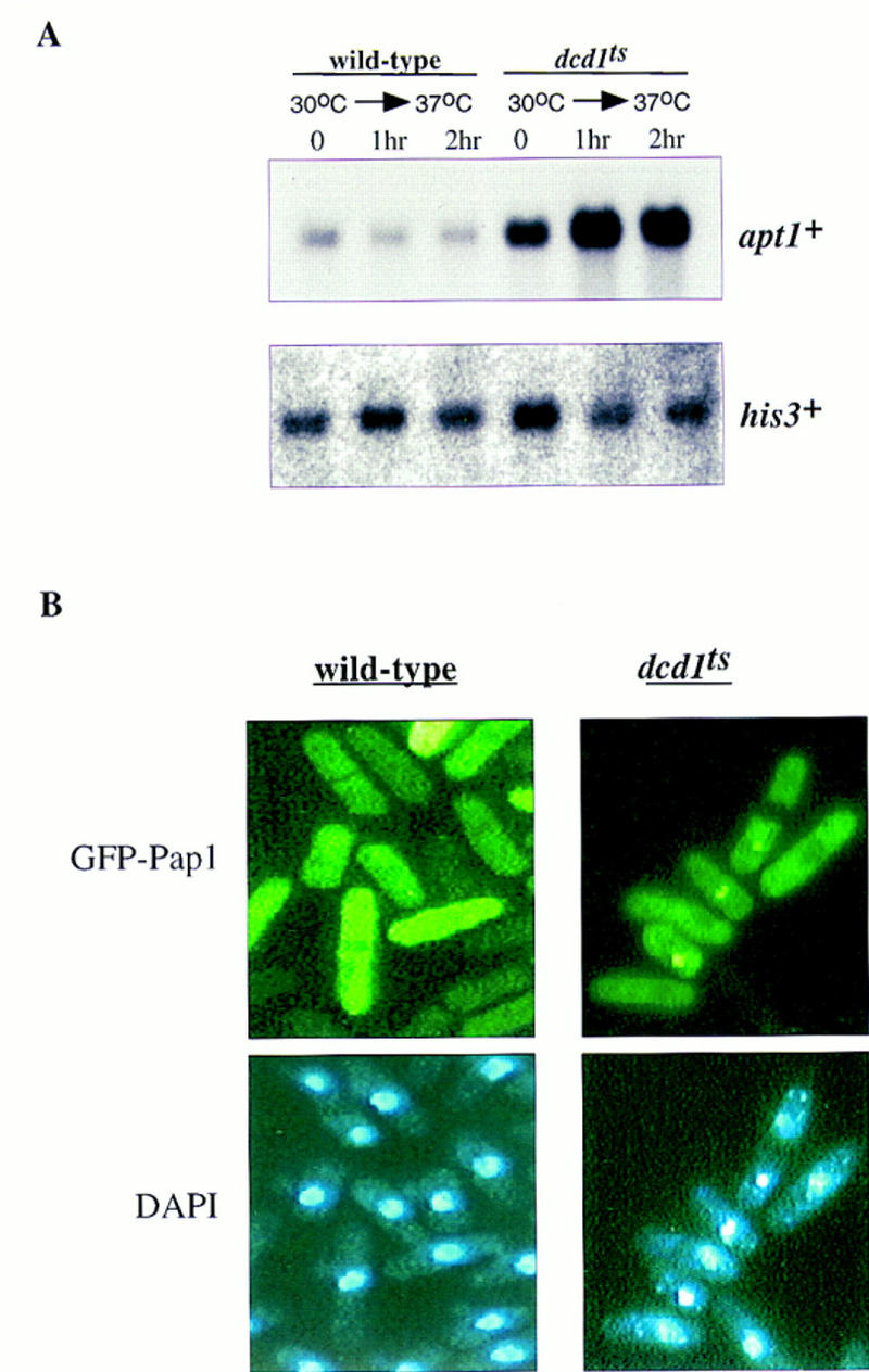
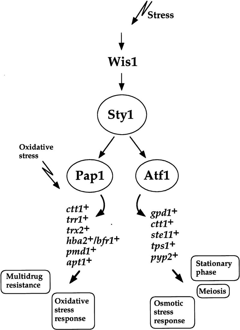
The fission yeast Sty1 stress-activated MAP kinase is crucial for the cellular response to a variety of stress conditions. Accordingly, sty1- cells are defective in their response to nutrient limitation, lose viability in stationary phase, and are hypersensitive to osmotic stress, oxidative stress, and UV treatment. Some of these phenotypes are caused by Sty1-dependent regulation of the Atf1 transcription factor, which controls both meiosis-specific and osmotic stress-responsive genes. However, in this report we demonstrate that the cellular response to oxidative stress and to treatment with a variety of cytotoxic agents is the result of Sty1 regulation of the Pap1 transcription factor, a bZip protein with structural and DNA binding similarities to the mammalian c-Jun protein. We show that both Sty1 and Pap1 are required for the expression of a number of genes involved in the oxidative stress response and for the expression of two genes, hba2+/bfr1+ and pmd1+, which encode energy-dependent transport proteins involved in multidrug resistance. Furthermore, we demonstrate that Pap1 is regulated by stress-dependent changes in subcellular localization. On imposition of oxidative stress, the Pap1 protein relocalizes from the cytoplasm to the nucleus in a process that is dependent on the Sty1 kinase. This relocalization is the result of regulated protein export, rather than import, and involves the Crm1 (exportin) nuclear export factor and the dcd1+/pim1+ gene that encodes an Ran nucleotide exchange factor.


Figures

Figure 1
Figure 1
Phenotypes of strains carrying mutations in pap1+, sty1+, or atf1+. pap1 and sty1 cells were hypersensitive to a variety of cytotoxic compounds. Approximately 103 cells from exponentially growing cultures of each strain were spotted onto YE5S plates containing the indicated compounds: t-BOOH (0.5 mm); diamide (2 mm); cadmium sulfate (0.1 mm); sodium arsenite (0.5 mm); cisplatin (0.5 mm); cycloheximide (15 μg/ml); staurosporine (0.5 μg/ml); anisomycin (7.5 μg/ml). Plates were incubated at 30°C for 2–3 days.
Figure 2
Figure 2
Northern analysis of stress-responsive genes in wild-type (wt), pap1, sty1, and atf1 strains following H2O2 treatment. (A) Total RNA was isolated from each of the indicated strains at 0, 20, and 60 min, following addition of H2O2 to a final concentration of 0.2 mm, electrophoresed, transferred to a nylon membrane, and probed with a 32P-labeled DNA specific to each of the genes denoted. The invariant his3+ mRNA was included as a loading control. (B) Total RNA was isolated from wild type (HM123) and HM123 carrying pRep1pap1+ Five micrograms of RNA was electrophoresed, transferred to a nylon membrane, and hybridized with 32P-labeled probes specific to either hba2+/bfr1+, pmd1+, or his3+. (C) Total RNA was isolated from the indicated strains before and after H2O2 treatment as described above, transfered to a nylon membrane, and probed with 32P-labeled probes specific to either hba2+/bfr1+, pmd1, or his3+.
Figure 2
Figure 2
Northern analysis of stress-responsive genes in wild-type (wt), pap1, sty1, and atf1 strains following H2O2 treatment. (A) Total RNA was isolated from each of the indicated strains at 0, 20, and 60 min, following addition of H2O2 to a final concentration of 0.2 mm, electrophoresed, transferred to a nylon membrane, and probed with a 32P-labeled DNA specific to each of the genes denoted. The invariant his3+ mRNA was included as a loading control. (B) Total RNA was isolated from wild type (HM123) and HM123 carrying pRep1pap1+ Five micrograms of RNA was electrophoresed, transferred to a nylon membrane, and hybridized with 32P-labeled probes specific to either hba2+/bfr1+, pmd1+, or his3+. (C) Total RNA was isolated from the indicated strains before and after H2O2 treatment as described above, transfered to a nylon membrane, and probed with 32P-labeled probes specific to either hba2+/bfr1+, pmd1, or his3+.
Figure 3
Figure 3
GFP–Pap1 localizes to the nucleus following oxidative stress. (A) A schematic of the GFP–pap1+ fusion construct expressed under the control of the intermediate strength nmt1+ promoter (pRep41). (B) Fluorescence microscopic analysis of untreated wild-type cells containing Rep41–GFP–Pap1, stained with DAPI to visualize the nucleus at 365 nm or at 390 nm to visualize the cellular distribution of GFP-tagged Pap1. (C) Fluorescence analysis of the same cells treated for 30 min with 0.2 mm H2O2.
Figure 4
Figure 4
Stress-dependent relocalization of GFP–Pap1 to the nucleus involves the Sty1 pathway. Wild-type cells (TP114-2A + pRep41–GFP–pap1+ + pRep42), sty1 cells (NT224 + pRep41–GFP–pap1+ + pRep42), or wild-type cells overexpressing the Wis1 kinase (TP114-2A + pRep41–GFP–pap1+ + pRep42–wis1+) were exposed to 0.2 mm H2O2 for the indicated times and subjected to fluorescence microscopy to visualize the GFP-tagged Pap1 fusion protein.
Figure 5
Figure 5
crm1 mutants show increased nuclear accumulation of GFP–Pap1. (A) Total RNA was isolated from exponentially growing cultures (30°C) of wild-type, crm1-809, or crm1-119 cells. Five micrograms of RNA was electrophoresed, transfered to a nylon (Genescreen) membrane and probed with a 32P-labeled probe specific to hba2+/bfr1+, apt1+, or his3+. (B) Exponentially growing wild-type cells, or crm1-809 cells, grown at 30°C, were subjected to fluoresence microscopy to determine the intracellular location of the GFP–Pap1 fusion protein.
Figure 6
Figure 6
RCC1 mutants show increased nuclear accumulation of GFP–Pap1. (A) Total RNA was isolated from exponentially growing cultures of wild-type cells and dcd1ts cells grown at 30°C or cells shifted to the nonpermissive temperature (36°C) for 1 or 2 hr. Five micrograms of RNA was electrophoresed, transferred to a nylon (Genescreen) membrane, and probed with a 32P-labeled probe specific to apt1+ and his3+ as a loading control. (B) Exponentially growing wild-type cells or dcd1ts cells were subjected to fluoresence microscopy to determine the intracellular location of the GFP–Pap1 fusion protein after a shift of cells from the permissive (30°C) temperature to 36°C for 2 hr.
Figure 7
Figure 7
Model depicting the role of the Sty1 pathway in the fission yeast stress response. The Sty1 signaling pathway is involved in the response of cells to a variety of different stress conditions. The role of Sty1 is to regulate the activity of the two transcription factors Atf1 (Shiozaki and Russell 1996; Wilkinson et al. 1996) and Pap1 (this study), which in turn regulate the expression of a number of genes encoding products that mediate different stress responses. The physiological events controlled by each factor are indicated.

Comment in

References

    1. Adachi Y, Yanagida M. Higher order chromosome structure is affected by cold-sensitive mutations in a Schizosaccharomyces pombe gene, crm1+, which encodes a 115-kDa protein preferentially localized to the nucleus and its periphery. J Cell Biol. 1989;108:1195–1207. - PMC - PubMed
    1. Alfa C, Fantes P, Hyams J, McLeod M, Warbrick E. Experiments with fission yeast. Cold Spring Harbor, N.Y: Cold Spring Harbor Laboratory Press; 1993.
    1. Baeuerle PA, Henkel T. Function and activation of NFκB in the immune system. Annu Rev Immunol. 1994;12:540–546. - PubMed
    1. Brewster JL, de Valoir T, Dwyer ND, Winter E, Gustin MC. An osmosensing signal transduction pathway in yeast. Science. 1993;259:1760–1763. - PubMed
    1. Casso D, Beach D. A mutation in a thioredoxin reductase homolog suppresses p53-induced growth-inhibition in the fission yeast Schizosaccharomyces pombe. Mol & Gen Genet. 1996;252:518–529. - PubMed

Publication types

MeSH terms

Substances