. 1998 May 15;12(10):1464-73.
doi: 10.1101/gad.12.10.1464.

Phosphorylation and association with the transcription factor Atf1 regulate localization of Spc1/Sty1 stress-activated kinase in fission yeast

Affiliations

Phosphorylation and association with the transcription factor Atf1 regulate localization of Spc1/Sty1 stress-activated kinase in fission yeast

F Gaits et al. Genes Dev. .

Abstract

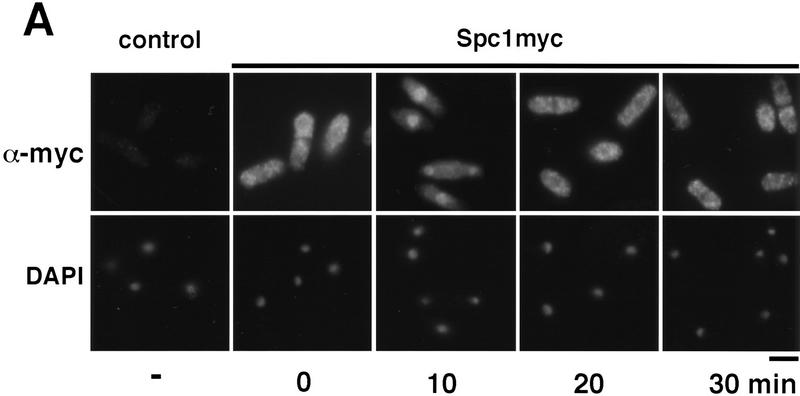
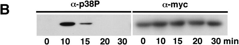
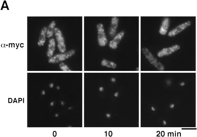
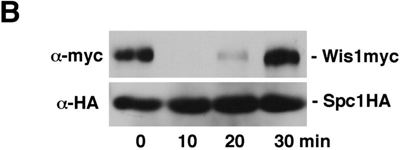
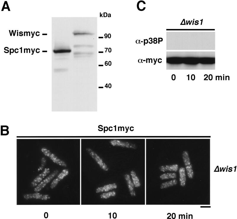
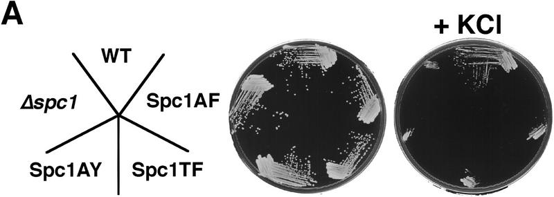
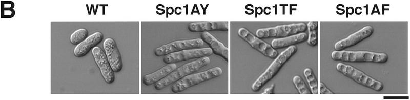
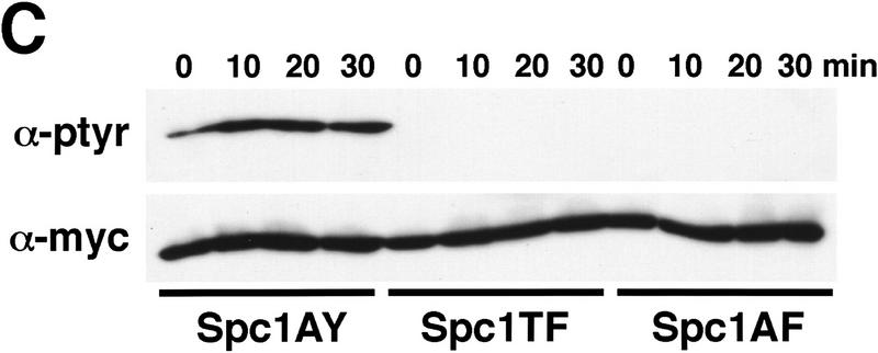
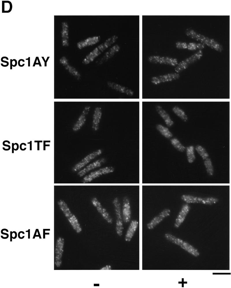
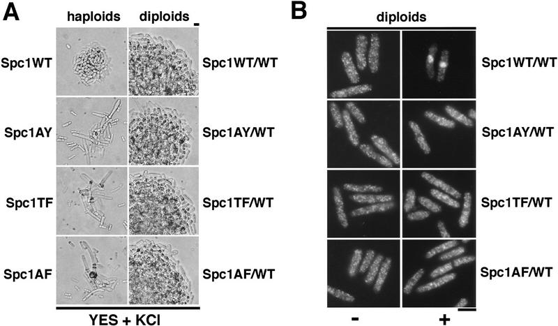
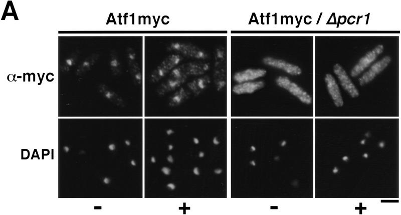
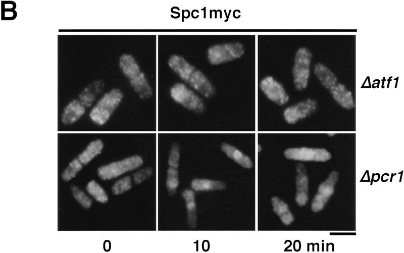
Control of gene expression by stress-activated protein kinase (SAPK) cascades is crucial for combating cytotoxic stress. Elements of these cascades have been investigated in detail, but regulation of stress signal transduction from the cytoplasm to the nucleus is poorly understood. Herein are reported subcellular localization studies of fission yeast Spc1, a homolog of human p38 and budding yeast Hog1p SAPKs. Stress induces transient nuclear localization of Spc1. Nuclear translocation of Spc1 is coupled with disassociation from its activator kinase Wis1. However, Spc1 does not concentrate in the nucleus of Deltawis1 cells; therefore Wis1 does not tether Spc1 in the cytoplasm. Unphosphorylatable forms of Spc1 are dispersed in the cytoplasm and nucleus, even in cells that also produce wild-type Spc1. Thus, Spc1 must be phosphorylated by Wis1 to localize in the nucleus. Nuclear retention of Spc1 requires Atf1, a transcription factor that is the key nuclear substrate of Spc1. Nuclear localization of Atf1 requires Pcr1, a heterodimerization partner of Atf1. These studies show that phosphorylation and association with Atf1 are required for nuclear localization of Spc1.


Figures

Figure 1
Figure 1
Spc1 localization with stress. (A) Spc1 translocates to the nucleus upon stress. Strain GD1942, in which the genomic copy of spc1+ encodes an epitope-tagged form of Spc1 that has 12 copies of a myc tag at the carboxyl terminus, was grown to mid-log phase at 30°C in YES medium. Aliquots were harvested before and after osmostress in YES + 0.6 m KCl at the indicated time and fixed in −80°C methanol. After permeabilization, cells were incubated with the anti-myc antibody and Cy3 goat anti-mouse as a secondary antibody to visualize Spc1myc. The nuclei were stained with DAPI. Bar, 10 μm. (B) In parallel, some aliquots were frozen in liquid nitrogen and total cellular homogenates were prepared to evaluate Spc1 phosphorylation. Proteins were subjected to SDS-PAGE and electroblotted, and immunodetection of Spc1 was achieved using the anti-p38 phosphorylated antibody (α-p38P). The level of proteins in each lane was evaluated using the anti-myc antibody (α-myc).
Figure 1
Figure 1
Spc1 localization with stress. (A) Spc1 translocates to the nucleus upon stress. Strain GD1942, in which the genomic copy of spc1+ encodes an epitope-tagged form of Spc1 that has 12 copies of a myc tag at the carboxyl terminus, was grown to mid-log phase at 30°C in YES medium. Aliquots were harvested before and after osmostress in YES + 0.6 m KCl at the indicated time and fixed in −80°C methanol. After permeabilization, cells were incubated with the anti-myc antibody and Cy3 goat anti-mouse as a secondary antibody to visualize Spc1myc. The nuclei were stained with DAPI. Bar, 10 μm. (B) In parallel, some aliquots were frozen in liquid nitrogen and total cellular homogenates were prepared to evaluate Spc1 phosphorylation. Proteins were subjected to SDS-PAGE and electroblotted, and immunodetection of Spc1 was achieved using the anti-p38 phosphorylated antibody (α-p38P). The level of proteins in each lane was evaluated using the anti-myc antibody (α-myc).
Figure 2
Figure 2
(A) Localization of Wis1 MAPKK. Strain GD1892, bearing a genomic copy of wis1+ tagged with 12 copies of the myc epitope at the carboxyl terminus, was grown to mid-log phase at 30°C in YES medium. Cells were harvested before and after stress in YES + 0.6 m KCl at the indicated time and fixed in cold methanol. Wis1 was visualized by immunofluorescence using anti-myc antibody. Bar, 10 μm. (B) Strain FG2150 bearing a genomic copy of spc1+ tagged with two copies of the HA-epitope and six consecutive histidine residues on its carboxyl terminus, as well as a genomic copy of wis1+ tagged with 12 copies of the myc epitope, was grown to mid-log phase and harvested before and after KCl stress at the indicated times. Total cell homogenates were then prepared under native conditions, and Spc1 was purified using Ni2+–NTA–agarose beads. The precipitates were analyzed by immunoblotting after SDS-PAGE. Wis1 was detected with the anti-myc and Spc1 with anti-HA epitope antibodies.
Figure 2
Figure 2
(A) Localization of Wis1 MAPKK. Strain GD1892, bearing a genomic copy of wis1+ tagged with 12 copies of the myc epitope at the carboxyl terminus, was grown to mid-log phase at 30°C in YES medium. Cells were harvested before and after stress in YES + 0.6 m KCl at the indicated time and fixed in cold methanol. Wis1 was visualized by immunofluorescence using anti-myc antibody. Bar, 10 μm. (B) Strain FG2150 bearing a genomic copy of spc1+ tagged with two copies of the HA-epitope and six consecutive histidine residues on its carboxyl terminus, as well as a genomic copy of wis1+ tagged with 12 copies of the myc epitope, was grown to mid-log phase and harvested before and after KCl stress at the indicated times. Total cell homogenates were then prepared under native conditions, and Spc1 was purified using Ni2+–NTA–agarose beads. The precipitates were analyzed by immunoblotting after SDS-PAGE. Wis1 was detected with the anti-myc and Spc1 with anti-HA epitope antibodies.
Figure 3
Figure 3
Wis1 activity is necessary to translocate Spc1 to the nucleus. (A) Spc1 is more abundant than Wis1. Total lysate of strains bearing myc-tagged genomic copies of Spc1 or Wis1, respectively, strains GD1942 and GD1892, were prepared and proteins separated by SDS-PAGE. The levels of Spc1 and Wis1 in the extract were analyzed by immunoblotting with the anti-myc antibody, the loading control being realized by amidoblack staining of the membrane prior to blotting. (B) Strain FG2151 (bearing the myc-tagged genomic copy of spc1+ and deleted for wis1) was grown to mid-log phase in YES medium and cells were harvested before and after stress with 0.6 m KCl. They were fixed in cold methanol, and Spc1 localization was examined by immunofluorescence using the anti-myc antibody. Bar, 10 μm. (C) Aliquots of cells were collected before and after KCl stress, and the phosphorylation status of Spc1 was analyzed by immunoblotting with the anti-p38P after SDS-PAGE. The loading control was realized by probing the membranes with anti-myc antibody.
Figure 4
Figure 4
Spc1 phosphorylation mutants are sensitive to high osmolarity and unable to relocate to the nucleus upon osmotic stress. (A) Strains FG2153 (genomic spc1+ replaced by spc1T171Amyc), FG2154 (genomic spc1+ replaced by spc1Y173Fmyc), FG2155 (genomic spc1+ replaced by spc1T171A Y173Fmyc), KS1366 (Δspc1), and PR109 (wild type) were streaked out onto YES agar plates supplemented with or without 1 m KCl, and incubated for 4 days at 32°C to test their ability to form colonies. (B) FG2153, FG2154, FG2155, and PR109 cells were streaked out on EMM2 agar plates. The phenotype of the cells was then analyzed by phase contrast microscopy after 2 days at 32°C. Bar, 10 μm. (C) The Spc1 mutant strains FG2153, FG2154 and FG2155 were grown to mid-log phase in YES medium and stressed with 0.6 m KCl before being harvested and frozen in liquid nitrogen. Total lysates were then prepared, and the phosphorylation status of the Spc1 mutants was analyzed by immunoblotting after SDS-PAGE using the anti-phosphotyrosine antibody (α-pTyr). The loading was checked using the anti-myc antibody (α-myc). (D) Strains FG2153 (Spc1AY), FG2154 (Spc1TF), FG2155 (Spc1AF) were grown to mid-log phase in YES medium, harvested before or after 10 min of incubation in YES supplemented by 0.6 m KCl, and fixed in cold methanol. The cellular localization of the mutant Spc1 was followed by indirect immunofluorescence with the anti-myc antibody. Bar, 10 μm.
Figure 4
Figure 4
Spc1 phosphorylation mutants are sensitive to high osmolarity and unable to relocate to the nucleus upon osmotic stress. (A) Strains FG2153 (genomic spc1+ replaced by spc1T171Amyc), FG2154 (genomic spc1+ replaced by spc1Y173Fmyc), FG2155 (genomic spc1+ replaced by spc1T171A Y173Fmyc), KS1366 (Δspc1), and PR109 (wild type) were streaked out onto YES agar plates supplemented with or without 1 m KCl, and incubated for 4 days at 32°C to test their ability to form colonies. (B) FG2153, FG2154, FG2155, and PR109 cells were streaked out on EMM2 agar plates. The phenotype of the cells was then analyzed by phase contrast microscopy after 2 days at 32°C. Bar, 10 μm. (C) The Spc1 mutant strains FG2153, FG2154 and FG2155 were grown to mid-log phase in YES medium and stressed with 0.6 m KCl before being harvested and frozen in liquid nitrogen. Total lysates were then prepared, and the phosphorylation status of the Spc1 mutants was analyzed by immunoblotting after SDS-PAGE using the anti-phosphotyrosine antibody (α-pTyr). The loading was checked using the anti-myc antibody (α-myc). (D) Strains FG2153 (Spc1AY), FG2154 (Spc1TF), FG2155 (Spc1AF) were grown to mid-log phase in YES medium, harvested before or after 10 min of incubation in YES supplemented by 0.6 m KCl, and fixed in cold methanol. The cellular localization of the mutant Spc1 was followed by indirect immunofluorescence with the anti-myc antibody. Bar, 10 μm.
Figure 5
Figure 5
Restoration of Spc1 activity does not rescue the localization defect of the unphosphorylatable mutants. (A) The haploid strains FG2153 (Spc1AY), FG2154 (Spc1TF), and FG2155 (Spc1AF), and the diploid strains FG2201 (Spc1WT/WT), FG2202 (Spc1AY/WT), FG2203 (Spc1TF/WT), and FG2204 (Spc1AF/WT) were streaked out onto YES agar plates supplemented with 1 m KCl and incubated for 3 days at 32°C. The phenotype of the cells was then analyzed by phase-contrast microscopy. (B) The diploid strains FG2201 (Spc1WT/WT), FG2202 (Spc1AY/WT), FG2203 (Spc1TF/WT), and FG2204 (Spc1AF/WT) were grown to mid-log phase in YES medium. Cells were then harvested before and after 10 min of KCl stress and fixed with cold methanol. The localization of the Spc1 proteins was then determined as described previously. Bar, 10 μm.
Figure 6
Figure 6
Atf1 acts as a nuclear anchor for Spc1. (A) The localization of Atf1 was determined by immunofluorescence performed on strain FG2156 in which the genomic atf1+ is replaced by a myc-tagged copy, and in the FG2157 (Δpcr1 and bearing the genomic myc-tagged atf1+). Cells were harvested before and after 10 min of KCl stress and fixed in cold methanol. The immunofluorescence was realized using the anti-myc antibody. (B) The strain GD1952 (Δatf1 and bearing the genomic myc-tagged copy of spc1+) and FG2158 (Δpcr1 and bearing the myc-tagged genomic spc1+) were grown to mid-log phase, and cells were harvested before and after KCl stress. For each time point, an aliquot was fixed in cold methanol for immunofluorescence analysis. The localization of Spc1 was determined with the anti-myc antibody. (C) The phosphorylation status of Spc1 in strains GD1952 and FG2158 was followed with the anti-p38P and the loading checked with the anti-myc antibodies, as described previously. Bar, 10 μm.
Figure 6
Figure 6
Atf1 acts as a nuclear anchor for Spc1. (A) The localization of Atf1 was determined by immunofluorescence performed on strain FG2156 in which the genomic atf1+ is replaced by a myc-tagged copy, and in the FG2157 (Δpcr1 and bearing the genomic myc-tagged atf1+). Cells were harvested before and after 10 min of KCl stress and fixed in cold methanol. The immunofluorescence was realized using the anti-myc antibody. (B) The strain GD1952 (Δatf1 and bearing the genomic myc-tagged copy of spc1+) and FG2158 (Δpcr1 and bearing the myc-tagged genomic spc1+) were grown to mid-log phase, and cells were harvested before and after KCl stress. For each time point, an aliquot was fixed in cold methanol for immunofluorescence analysis. The localization of Spc1 was determined with the anti-myc antibody. (C) The phosphorylation status of Spc1 in strains GD1952 and FG2158 was followed with the anti-p38P and the loading checked with the anti-myc antibodies, as described previously. Bar, 10 μm.

Comment in

Similar articles

Cited by

References

    1. Alfa C, Fantes P, Hyams J, McLeod M, Warbrick E. Experiments with fission yeast. Cold Spring Harbor, NY: Cold Spring Harbor Laboratory Press; 1993.
    1. Basi G, Schmid E, Maundrell K. TATA box mutations in the Schizosaccharomyces pombe nmt1 promoter affect transcription efficiency but not the transcription start point or thiamine repressibility. Gene. 1993;123:131–136. - PubMed
    1. Canagarajah BJ, Khokhlatchev A, Cobb MH, Goldsmith EJ. Activation mechanism of the MAP kinase ERK2 by dual phosphorylation. Cell. 1997;90:859–869. - PubMed
    1. Cano E, Mahadevan LC. Parallel signal processing among mammalian MAPKs. Trends Biochem Sci. 1995;20:117–122. - PubMed
    1. Cavigelli M, Dolfi F, Claret FX, Karin M. Induction of c-fos expression through JNK-mediated TCF/Elk-1 phosphorylation. EMBO J. 1995;14:5957–5964. - PMC - PubMed

Publication types

MeSH terms

Substances

LinkOut - more resources