. 1998 May 15;12(10):1474-82.
doi: 10.1101/gad.12.10.1474.

Ultrabithorax regulates genes at several levels of the wing-patterning hierarchy to shape the development of the Drosophila haltere

Affiliations

Ultrabithorax regulates genes at several levels of the wing-patterning hierarchy to shape the development of the Drosophila haltere

S D Weatherbee et al. Genes Dev. .

Abstract

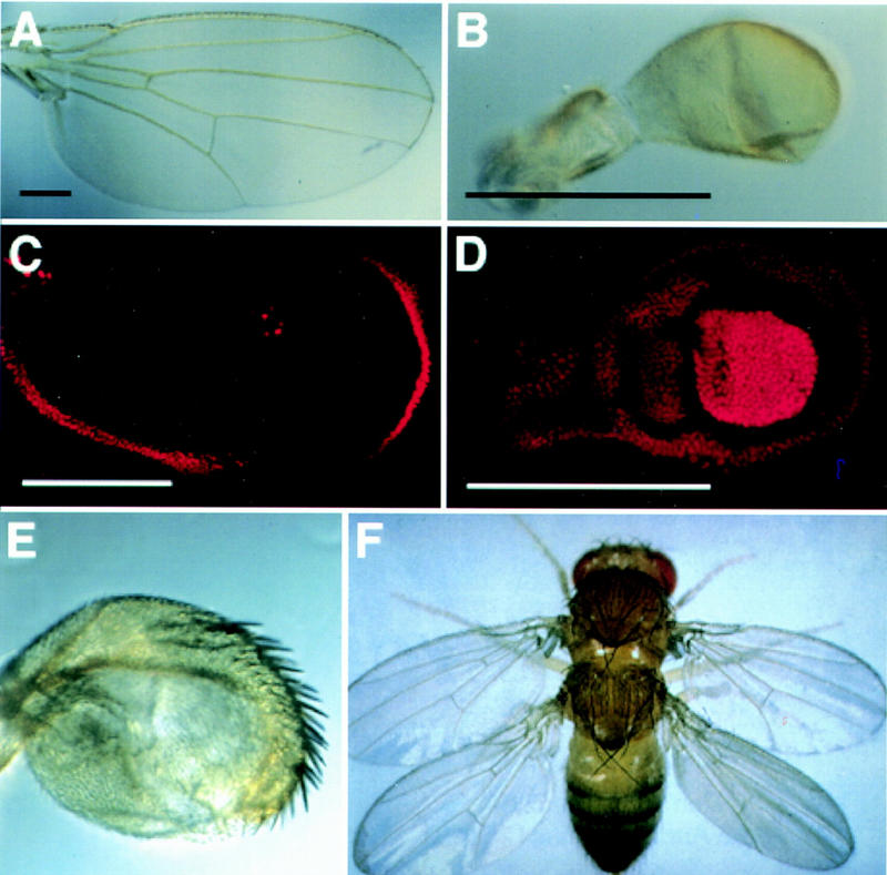
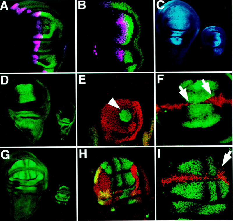
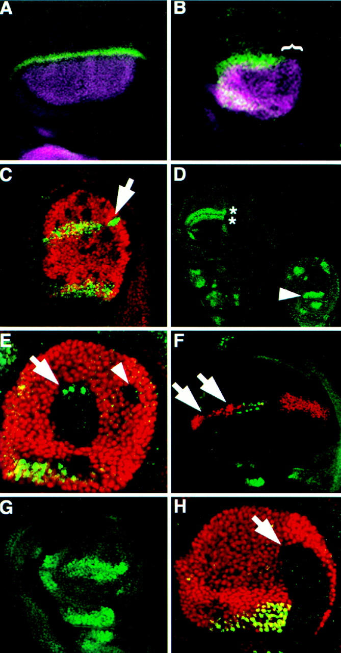
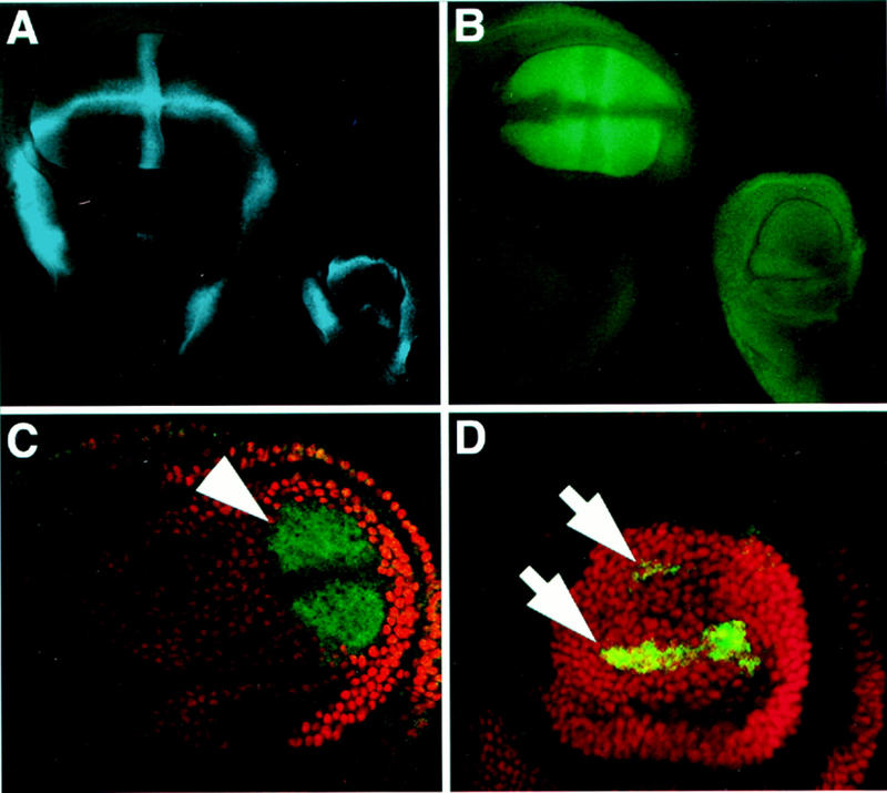
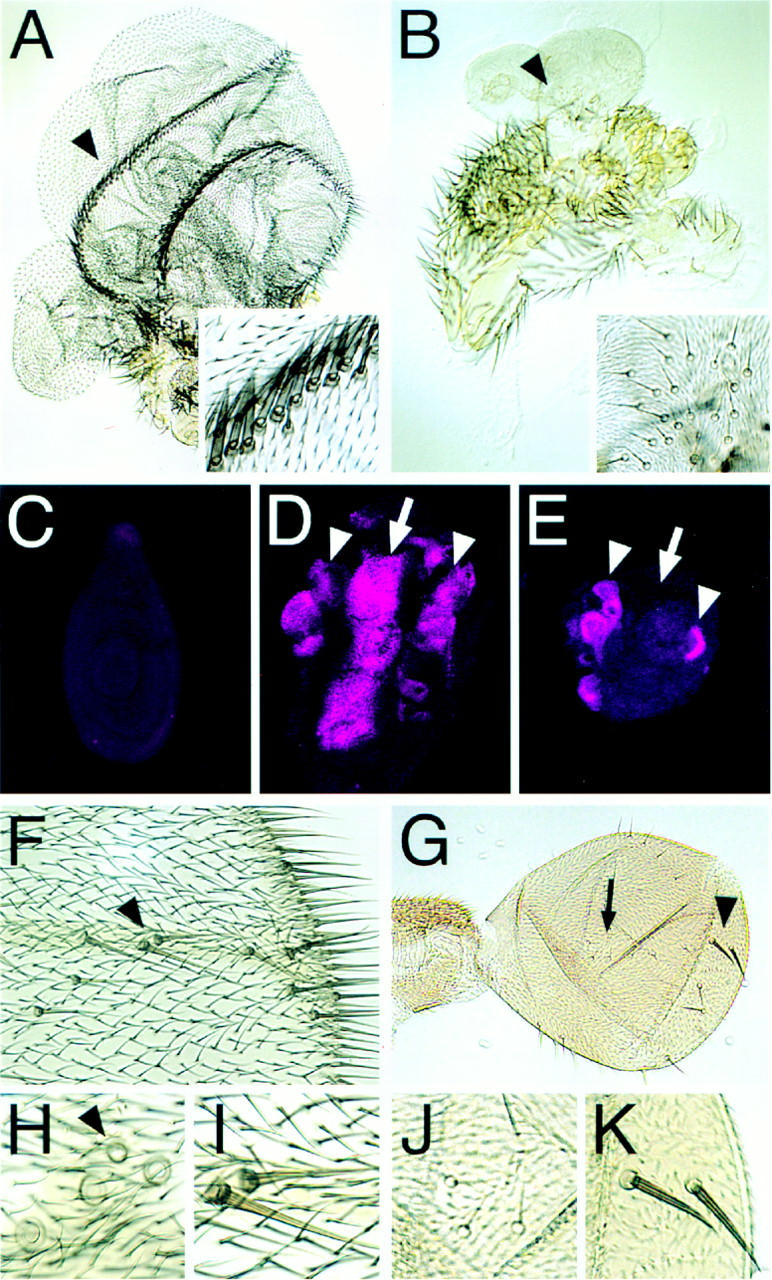
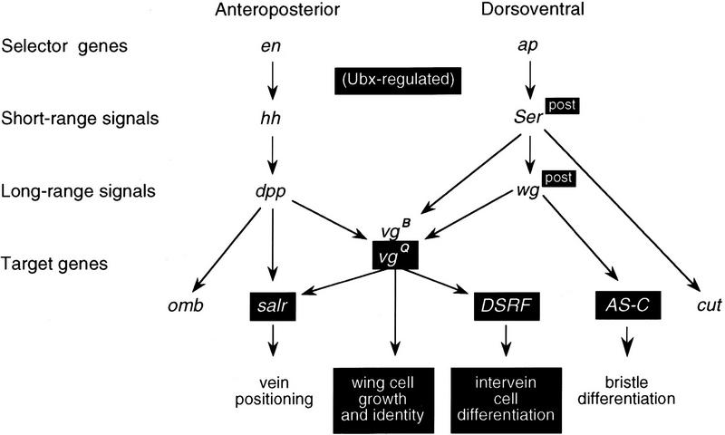
Arthropods and vertebrates are constructed of many serially homologous structures whose individual patterns are regulated by Hox genes. The Hox-regulated target genes and developmental pathways that determine the morphological differences between any homologous structures are not known. The differentiation of the Drosophila haltere from the wing through the action of the Ultrabithorax (Ubx) gene is a classic example of Hox regulation of serial homology, although no Ubx-regulated genes in the haltere have been identified previously. Here, we show that Ubx represses the expression of the Wingless (Wg) signaling protein and a subset of Wg- and Decapentaplegic-activated genes such as spalt-related, vestigial, Serum Response Factor, and achaete-scute, whose products regulate morphological features that differ between the wing and haltere. In addition, we found that some genes in the same developmental pathway are independently regulated by Ubx. Our results suggest that Ubx, and Hox genes in general, independently and selectively regulate genes that act at many levels of regulatory hierarchies to shape the differential development of serially homologous structures.


Figures

Figure 1
Figure 1
Ubx controls the differential development of the haltere. The wild-type wing (A) and haltere (B) differ in size, shape, and the presence of veins and margin bristles. (C,D) antibody staining of third instar wing and haltere discs. (C) Ubx expression (red) in the wing disc is limited to the peripodial membrane and is not necessary for proper wing development (Struhl 1982). (D) Ubx expression fills the haltere disc, with strongest expression in the “pouch”, which will give rise to capitellar tissue (Beachy et al. 1985). Reduction of Ubx activity in the halteres leads to transformations toward wing identity. (E) Haltere from a Ubx6.28/bx34E fly (Kerridge and Morata 1982), in which Ubx gene activity is <50% of wild-type (shown at the same magnification as B). A large number of ectopic margin bristles appear on the haltere, which is increased in size. (F) Total loss of Ubx activity in the developing haltere results in a complete transformation toward wing identity (Lewis 1978). Black scale bars, 0.25 mm; white scale bars, 0.2 mm.
Figure 2
Figure 2
Ubx represses genes downstream of AP patterning signals in the haltere. (A,B) En (green) and dpp (purple, visualized by a lacZ reporter transgene) expression patterns are similar in the wing (A) and haltere (B). (C) omb expression (blue, visualized by a lacZ reporter transgene) is found in the haltere disc (right) in a pattern similar to that found in the wing (left), indicating that Dpp signaling is not repressed by Ubx. (D–F) Antibody staining detecting Salr is shown in green; Ubx is shown in red. (D) Salr expression in a wing disc (left) and in a haltere disc (right). Salr is not expressed in the haltere pouch, indicating that this Dpp target gene is repressed by Ubx. (E) Ubx clone close to the AP boundary shows cell-autonomous derepression of Salr expression (arrowhead). Ubx clones more than eight cells anterior to the AP boundary and posterior clones do not show Salr derepression (not shown). (F) Ectopic Ubx expression in a CbxM1 heterozygous wing pouch represses Salr expression in a cell autonomous fashion (arrows). (G–I) antibody staining detecting DSRF is shown in green; Ubx is shown in red. (G, left), DSRF is expressed in the future intervein cells of the Drosophila wing imaginal disc. (right) Expression of DSRF in the haltere is limited to extreme ventral and dorsal crescents in the pouch, and is also present in pedicellar and notal portions of the disc. (H) Ubx clone in the haltere (lack of red staining) showing DSRF derepression in the haltere pouch in a cell-autonomous manner. A winglike pattern forms in the clone, whereas the haltere expression pattern is still visible where Ubx is expressed (yellow overlap). (I) Ectopic Ubx expression in a CbxM1/+ wing disc represses a portion (ventral intervein D) of the normal DSRF expression (arrw). Note that omb expression does not extend into the posterior of the haltere nearly as far as it does in the anterior (Fig. 2C), and Salr expression is not derepressed in posterior Ubx clones close to the AP boundary (not shown) which suggests that Dpp signaling may somehow be reduced in the posterior haltere disc.
Figure 3
Figure 3
Ubx represses selected genes along the DV boundary of the haltere disc. (A–C) Antibody staining detecting Wg (green); Ap (purple), Ubx (red). (A,B) Ap and Wg are expressed in a similar domain in the haltere (B) as in the wing (A), but Wg expression is absent from the posterior haltere (bracket). (C) Haltere disc with several Ubx clones (lack of red staining). A posterior clone, located along the DV boundary (arrow) shows derepression of Wg expression. (D–H) Antibody staining detecting Sc (green); Ubx (red). (D) Wild-type expression of Sc in the wing (left) and haltere (right) disc pouches. The double row of expression in sensory organ precursors along the future wing margin (asterisks) is absent from the haltere disc. In the haltere disc, Sc is also expressed in unique patterns including the pedicellular region (arrowhead). (E) Haltere disc with two dorsal Ubx clones that each touch the DV boundary. The anterior (arrow) clone shows derepression of Sc expression; the posterior (arrowhead) clone shows no Sc expression as in the posterior of the wing. (F) Ubx expression along the DV boundary of a CbxM1/+ wing disc represses Sc expression along the presumptive anterior wing margin (arrows). (G) Ubx6.28/bx34e haltere disc showing ectopic Sc expression along the anterior DV boundary. (H) Ubx clone (arrow) crossing into the pedicellar region of the haltere disc (see arrowhead in D) fails to activate the normal Sc expression there indicating that Ubx is necessary for activation of Sc in the pedicellar region of the haltere.
Figure 4
Figure 4
Ubx selectively regulates one enhancer of the vg gene. (A) The Notch-regulated vg boundary enhancer (blue, visualized by a lacZ reporter transgene) is activated along the DV boundary and hinge region in both the wing and haltere discs. (B–D) vg quadrant enhancer expression is visualized by lacZ (green) and antibody staining detecting Ubx (red). (B) vg quadrant enhancer expression fills the wing pouch (left) in a pattern complementary to the vg boundary enhancer, but is silent in the haltere (right). (C) Ubx6.28/+ haltere disc pouch showing derepression of the vg quadrant enhancer (arrows), indicating that repression of this enhancer is sensitive to Ubx gene dosage. (D) Ubx clone in a haltere disc showing derepression of the vg quadrant pattern (arrowhead) that extends to the clone borders. The decreased expression along the DV boundary in the center of the clone is a normal feature of quadrant enhancer expression in the wing disc (see B).
Figure 5
Figure 5
Targeted expression of Ubx-regulated genes. (A–E) Ectopic expression of Vg, (F–K) ectopic expression of Sc. (A) Transformation of a T2 leg to wing as a consequence of ectopic Vg expression. Two “wing margins” formed in this specimen showing the prominent triple row of margin bristles (region pointed out by arrowhead is magnified in inset). The hairs in the “blade region” are also of wing identity. (B) Transformation of a T3 leg towards haltere. Magnification is the same as in A. The ectopic structure is much smaller than that formed on the T2 leg and the hair morphology and density are like those found on a haltere. In addition, a cluster of what appears to be capitellar sensillae appear that are characteristic for the haltere (magnified in inset). (C) T2 leg imaginal disc of a wild-type third instar larva stained for Sal protein. No expression is detected. Sal is also not expressed in T1 and T3 leg discs (not shown). (D) Upon targeted expression of Vg, Sal is ectopically induced in T2 legs in a broad stripe along the AP axis (arrow) similar to its expression pattern in the developing wing pouch (compare to Fig. 2D). In addition, Sal is ectopically induced in smaller domains towards the periphery (proximal region) of the disc (arrowheads) that may correspond to Sal expression domains in the presumptive hinge and notal regions of the wing disc. (E) In contrast to the T2 leg disc, in T3 leg discs, ectopic Vg is unable to induce Sal expression along the AP axis. The corresponding pattern is normally repressed by Ubx in the developing haltere. Therefore, targeted expression of Vg is unable to override the Ubx-regulation of the downstream gene sal. (F) Distal part of a wing. Anterior is to the top, distal to the right. Extra bristles, resembling double row margin bristles developed along the distal AP boundary. The two bristles marked by the arrowhead are magnified in I. (H) More proximlly on the wing, mainly campaniform sensillae are induced. The region of the anterior crossvein where four instead of only one (arrowhead) campaniform sensilla developed. (G) Haltere; same orientation and magnification as F. On the proximal haltere, ectopic capitellar sensillae are induced (arrow, magnified in J); on the distal haltere, ectopic bristles resembling double row wing margin bristles developed (arrowhead, magnified in K).
Figure 6
Figure 6
The architecture of the Ubx-regulated gene hierarchy in the haltere. The products of the Ap and En selector genes and the Hh and Dpp signaling proteins are expressed similarly in the wing and haltere. Ser and Wg are expressed similarly in the anterior of the wing and haltere but not in the posterior. A selected subset of the target genes activated by Dpp, Wg, or other pathways are regulated by Ubx (shown boxed). Note that some target genes are also upstream activators or coactivators of genes which are also Ubx-regulated. The Salr, Vg, Sc, and DSRF products in turn affect the differentiation of veins, wing and haltere cells, sensory organs, and intervein cells. Ubx regulation can operate selectively upon different enhancers of the same gene. The vg quadrant enhancer (vgQ) is Ubx-regulated; the vg boundary enhancer (vgB) is not.

References

    1. Beachy PA, Helfand SL, Hogness DS. Segmental distribution of bithorax complex proteins during Drosophila development. Nature. 1985;313:545–551. - PubMed
    1. Brand A, Perrimon N. Targeted gene expression as a means of altering cell fates and generating dominant phenotypes. Development. 1993;118:401–415. - PubMed
    1. Burke R, Basler K. Hedgehog signaling in Drosophila eye and limb development-conserved machinery, divergent roles? Curr Opin Neurobiol. 1997;7:55–61. - PubMed
    1. Cabrera C, Botas J, Garcia-Bellido A. Distribution of Ultrabithorax proteins in mutants of Drosophila bithorax complex and its transregulatory genes. Nature. 1985;318:569–571.
    1. Carroll S. Homeotic genes and the evolution of arthropods and chordates. Review. Nature. 1995;376:479–485. - PubMed

Publication types

MeSH terms

LinkOut - more resources