Comparative Study
. 1998 May 15;12(10):1483-94.
doi: 10.1101/gad.12.10.1483.

pavarotti encodes a kinesin-like protein required to organize the central spindle and contractile ring for cytokinesis

Affiliations
Comparative Study

pavarotti encodes a kinesin-like protein required to organize the central spindle and contractile ring for cytokinesis

R R Adams et al. Genes Dev. .

Abstract

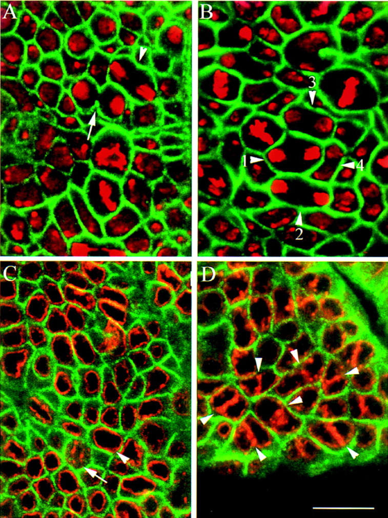
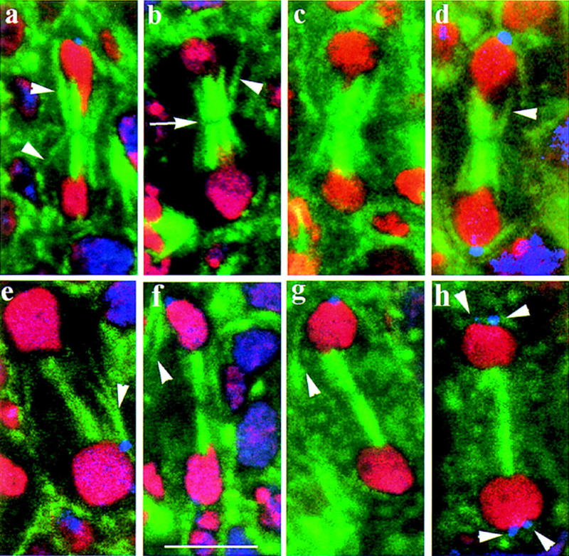
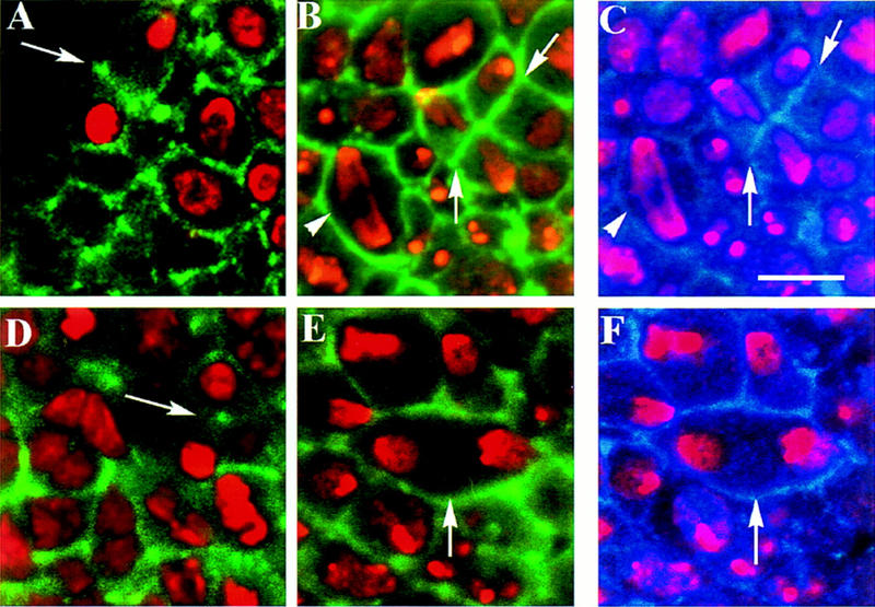
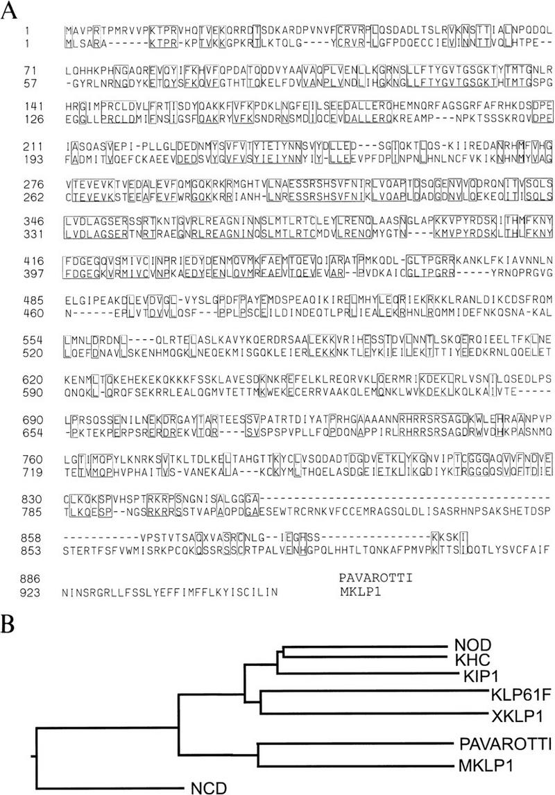
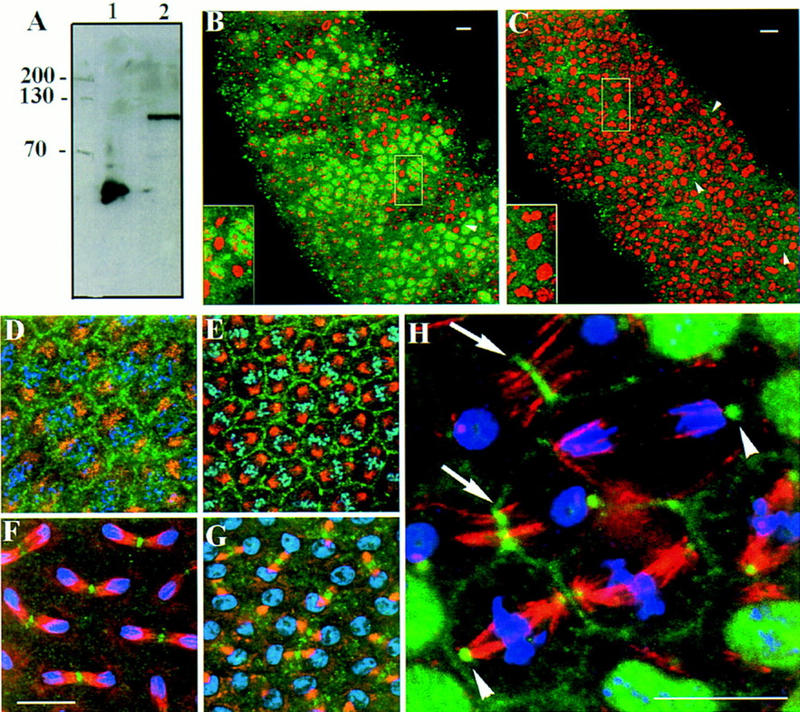
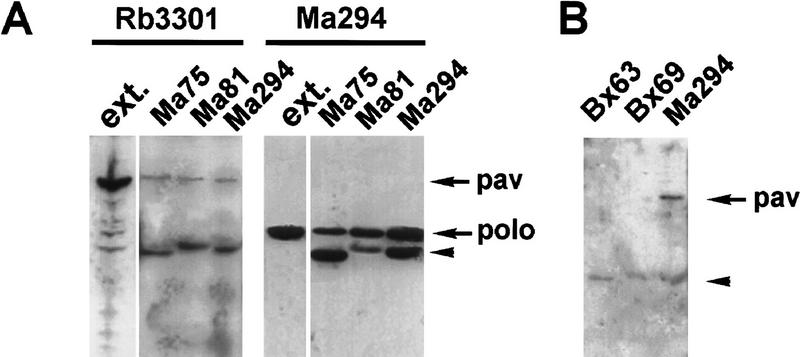
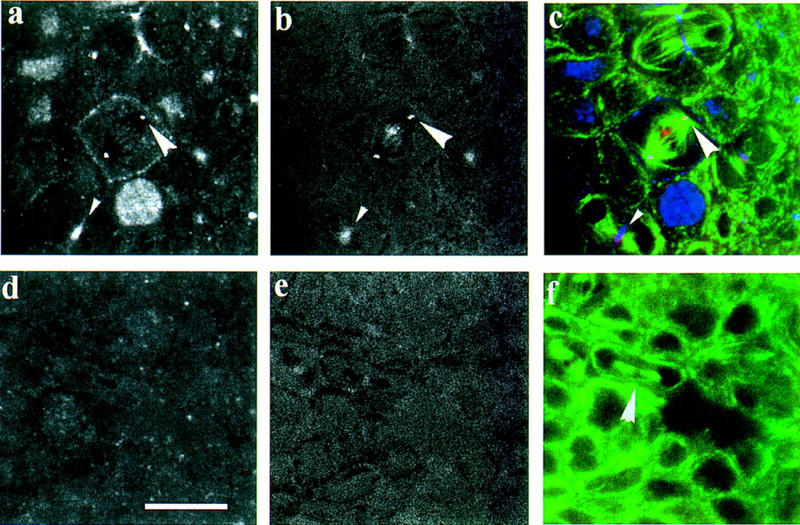
Mutations in the Drosophila gene pavarotti result in the formation of abnormally large cells in the embryonic nervous system. In mitotic cycle 16, cells of pav mutant embryos undergo normal anaphase but then develop an abnormal telophase spindle and fail to undertake cytokinesis. We show that the septin Peanut, actin, and the actin-associated protein Anillin, do not become correctly localized in pav mutants. pav encodes a kinesin-like protein, PAV-KLP, related to the mammalian MKLP-1. In cellularized embryos, the protein is localized to centrosomes early in mitosis, and to the midbody region of the spindle in late anaphase and telophase. We show that Polo kinase associates with PAV-KLP with which it shows an overlapping pattern of subcellular localization during the mitotic cycle and this distribution is disrupted in pav mutants. We suggest that PAV-KLP is required both to establish the structure of the telophase spindle to provide a framework for the assembly of the contractile ring, and to mobilize mitotic regulator proteins.


Figures

Figure 1
Figure 1
pav embryos show cytokinesis defects at cycle 16. (A) A wild-type embryo at cycle 16 stained to reveal α-spectrin (green) and DNA (red). The contraction of the cleavage furrow can be seen in cells in late anaphase (arrowhead) and telophase (arrow). (B) Cells in homozygous pavB200 embryos do not develop this constriction in cycle 16. Arrowheads 1–4 show the development of binucleate cells at progressively later stages from anaphase through telophase. (C) A wild-type embryo at cycle 16 stained to reveal lamin A (red) and α-spectrin (green). There is only one nuclear lamina per cell (arrowhead), which is dismantled during mitosis (arrow). (D) Homozygous pavB200 embryo at cycle 16, with many cells containing two nuclear laminae (arrowheads). Scale bar, 10 μm.
Figure 2
Figure 2
pav embryos show abnormal central spindles. A selection of confocal images showing telophase spindles from cycle 16 wild type (a–d) and pav (e–h) embryos stained to reveal β-tubulin (green), DNA (red), and the centrosomal antigen CP190 (blue). The wild-type spindle at telophase comprises a dense network of microtubules extending between the separated nuclei. Peripheral microtubules taper inward at the spindle equator due to the action of the contractile ring (arrowheads in a–d). A clear gap between the two halves of the spindle is also apparent (arrow in b). In contrast, the peripheral microtubules in pav telophases fail to taper inward (arrowheads in e–g) and the microtubules of the central spindle are generally more compact (a–h). (h) A rare telophase cell with four centrosomes and large nuclei. Measurements of centrosome–centrosome distances at metaphase and anaphase revealed no differences between wild type and pav mutants. Spindle lengths were 5.46 ± 0.46 μm (n = 32) wild-type metaphase; 9.41 ± 1.00 μm (n = 43) wild-type anaphase; 5.43 ± 0.41 μm (n = 10) pav metaphase; 10.22 ± 0.96 μm (n = 17) pav anaphase. Scale bar, 10 μm.
Figure 3
Figure 3
Neither Peanut protein, Actin, or Anillin becomes correctly localized at the end of mitosis in pav embryos. Wild-type (A-C) and pav (D–F) cycle 16 embryos were stained to reveal DNA (red) and either Peanut (A and D, green), Actin (B and E, green), or Anillin (C and F, blue). In wild type (A), Peanut is distributed beneath the cell surface, and at anaphase it accumulates near the nascent contractile ring (arrow) and remains associated with this region through telophase (arrow). In pav embryos (D) Peanut protein is variable in distribution and more diffuse than in wild type. Peanut is notably absent from the equatorial region of the cell during telophase (arrow). Dividing cells from wild-type embryos (B and C) accumulate actin (B) in the equatorial region at late anaphase (short arrow) through telophase (long arrow). The arrowhead shows a mid-anaphase figure, before this accumulation has occurred. The distribution of Anillin (C) at the end of mitosis is very similar to that of Actin. In addition, anillin shows faint nuclear staining during interphase. In pav embryos, neither Actin nor Anillin localize to the equatorial region during telophase (arrows in E and F, respectively). Scale bar, 10 μm.
Figure 4
Figure 4
Physical map of the pav region. (A) Genomic fragments obtained by plasmid rescue of P-element mutants. (B) Restriction endonuclease cleavage map of a 20.5-kb segment of genomic DNA. Cleavage sites are: EcoRI (R); XbaI (X); StuI (St); PstI (P); HindIII (H); SmaI (Sm). (C) Location and polarity of 3.2-kb pNBpav cDNA. (D) Transformation constructs to test rescue of pav phenotype.
Figure 5
Figure 5
Comparison of the PAV–KLP with MKLP-1. (a) The full nucleotide sequence of the pNBpav cDNA and corresponding genomic region have been deposited in the EMBL databank. There are five putative initiation ATGs between the site of the P-element insertion and the longest ORF present in pNBpav. This ORF, located 191 bp, 3′ to the site of the P-element insertion identifies a protein of 886 amino acids with extensive homology to MKLP-1 and a predicted molecular mass of 100 kD. PAV–KLP contains a consensus nuclear localization motif (KTPR) at amino acids 13–17, an ATP binding site (GSGKT) at amino acids 132–137, and two motifs common to all KLPs (SSRSHS and LAGSE) at residues 308–313 and 349–352, respectively. Analysis of the sequence using the Lupas algorithm (Lupas et al. 1991) predicts the formation of coiled–coil regions in the central region of the protein (residues 500–700; data not shown). The comparison of the amino acid sequences of PAV–KLP (top row) and MKLP-1 (bottom row) was made by Clustal analysis using DNASTAR software (Lasergene). Identical residues are boxed. Both sequences show substantial sequence identity in the first 450 amino acids, corresponding to the conserved motor domain. Regions of sequence identity also extend into the carboxy-terminal domains. Overall, PAV-KLP shows 34.3% identity with MKLP-1. (B) A phylogenetic tree illustrating the sequence homology between mitotic KLPs. PAV–KLP is related more closely to MKLP-1 than any other KLP shown. Other than these two KLPs, the remaining members of this superfamily that are shown are Drosophila NOD; Drosophila KHC (kinesin heavy chain); S. cerevisiae KIP1; Drosophila KLP61F; Xenopus XKLP1, and Drosophila NCD (Yang et al. 1988; McDonald et al. 1990; Zhang et al. 1990; Hoyt et al. 1992; Heck et al. 1993; Vernos et al. 1995).
Figure 6
Figure 6
Subcellular localization of PAV–KLP at division. (A) Western blot analysis of E. coli cells induced to express the construct PpavEC2, and 0- to 4-hr Drosophila embryo extracts probed with the rabbit antiserum Rb3301. The antiserum recognizes the 52-kD polypeptide against which it was raised (lane 1) and a single protein of ∼100 kD in the embryo extracts (lane 2). We were unable to carry out Western analysis on homozygous mutant embryos, as it proved difficult to distinguish these from their heterozygous siblings on the basis of the phenotype revealed by DNA staining alone. We could distinguish homozygotes from heterozygotes carrying a balancer chromosome expressing lacZ, but neither immuno- nor histochemical staining methods to detect β-galactosidase preserved proteins sufficiently well for Western analysis. (B) Homozygotes and heterozygotes can, however, be distinguished by immunostaining with the anti-pav antibody, even though this antibody is not sensitive enough to detect levels of protein from single embryos on Western blots. TM6B/TM6B or pavB200/TM6B embryos show strong nuclear staining in segmented bands of interphase cells using the antibody Rb3301 (green; DNA staining is in red). Bright regions of staining are also seen equidistant from separating anaphase cells (arrowhead and inset). Smaller punctate staining is also visible scattered throughout the embryo. (C) pavB200 homozygotes show neither nuclear staining at interphase nor equatorial staining at anaphase using the Rb3301 antibody (arrowheads and inset). However, there are some spots of staining distributed throughout the embryo, likely to correspond to the staining of residual maternally contributed protein. (D–G) Syncytial wild-type type embryos were stained to reveal PAV–KLP with Rb3301 (green), tubulin with monoclonal antibody YL1/2 (red), and DNA with TOTO-3 (blue). At prophase (D), PAV–KLP is diffusely distributed but is beginning to locate to the furrows separating the mitotic nuclei. PAV–KLP localization to the furrows persists through metaphase (E). Upon initiation of anaphase (F), PAV–KLP relocates to the spindle interzone region. At late telophase (G) PAV–KLP staining is concentrated at the midbody region of the spindle, and diffuse staining is reappearing in the cytoplasm. (H) A cellularized embryo at cycle 14 showing cells at various stages of mitosis. PAV–KLP is visible at the centrosomes in metaphase and anaphase cells (arrowheads) but does not appear at the spindle midzone until telophase, where it clearly colocalizes with microtubules (arrows). Interphase cells show punctate nuclear staining.
Figure 7
Figure 7
PAV–KLP coimmunoprecipitates with Polo protein kinase. (A) Polo was immunoprecipitated from Drosophila embryo extracts using three different antibodies (mAb 75, mAb 81, mAb 294), and the immunoprecipitates were analyzed by Western blot as described in Materials and Methods. An aliquot of the embryo extract was loaded in a separate lane as a control (ext.). The Western blot was first probed with mAb Rb3301 to detect PAV–KLP, and after washing, the membrane was reprobed with mAb 294 to detect Polo protein. (B) A comparison of immunoprecipitates obtained using Bx63, a mAb against the centrosomal-associated protein CP190; an anti-β-tubulin mAb Bx69; and the anti-polo mAb Ma294. The Western blot is probed with the anti-PAV-KLP Rb3301. (A,B) The lower band marked by the arrowhead corresponds to the antibodies used for the immunoprecipitations that are detected by the secondary antibody used in the Western blot.
Figure 8
Figure 8
The colocalization of Polo kinase with PAV–KLP at centrosomes and in the central spindle is disrupted in pav mutant embryos. Wild-type (a–c) or pavB200 (d–f) cycle 16 embryos stained for either PAV–KLP (a,d) or Polo (b,e). (e,f) Merged images of PAV–KLP (blue), Polo (red), and tubulin (green). In wild type, Polo colocalizes with PAV–KLP at the centrosome (large arrowhead in a–c) and the midbody (small arrowhead in a–c). In addition, faint Polo staining is apparent at the metaphase plate. However, in pav embryos, neither PAV–KLP (d) nor Polo (e) is present at the centrosomes or midbody of a mutant telophase (arrowhead in f).

References

    1. Andreassen PR, Palmer DK, Wener MH, Margolis RL. Telophase disc: A new mammalian mitotic organelle that bisects telophase cells with a possible function in cytokinesis. J Cell Sci. 1991;99:523–534. - PubMed
    1. Bonaccorsi S, Giansanti MG, Gatti M. Asterless, a Drosophila gene specifically required for the formation of asters during male meiosis. Mol Biol Cell. 1996;7S:574a.
    1. Brown NH, Kafatos FC. Functional cDNA libraries from Drosophila embryos. J Mol Biol. 1988;203:425–437. - PubMed
    1. Burton K, Taylor DL. Traction forces of cytokinesis measured with optically modified elastic substrata. Nature. 1997;385:450–454. - PubMed
    1. Cao L-G, Wang Y-L. Signals from the spindle midzone are required for the stimulation of cytokinesis in cultured epithelial cells. Mol Biol Cell. 1996;7:225–232. - PMC - PubMed

Publication types

MeSH terms

LinkOut - more resources