Comparative Study
. 1998 May 15;12(10):1525-38.
doi: 10.1101/gad.12.10.1525.

Spalten, a protein containing Galpha-protein-like and PP2C domains, is essential for cell-type differentiation in Dictyostelium

Affiliations
Comparative Study

Spalten, a protein containing Galpha-protein-like and PP2C domains, is essential for cell-type differentiation in Dictyostelium

L Aubry et al. Genes Dev. .

Abstract

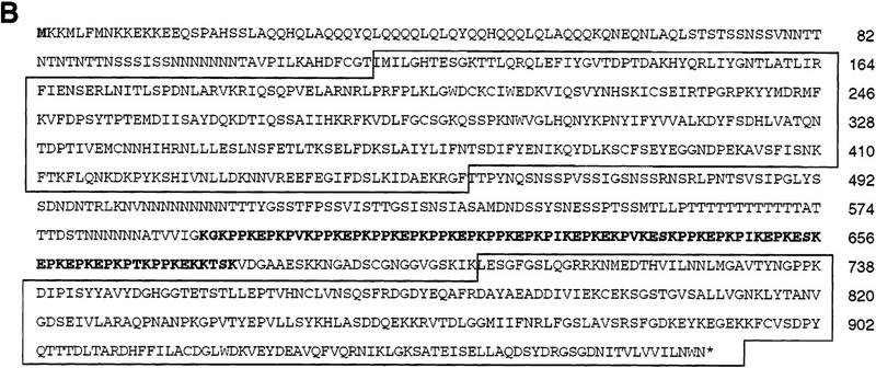
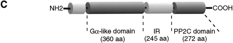
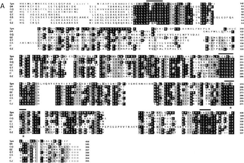
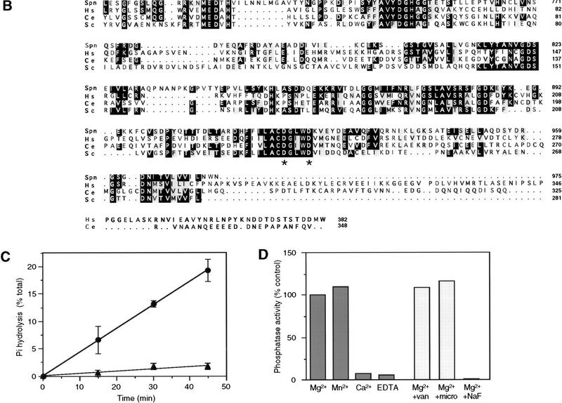
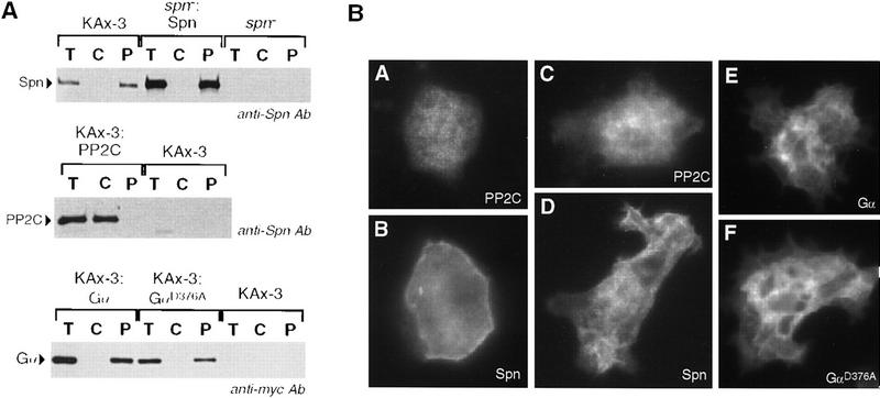
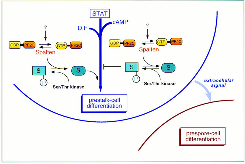
We have identified a novel gene, Spalten (Spn) that is essential for Dictyostelium multicellular development. Spn encodes a protein with an amino-terminal domain that shows very high homology to Galpha-protein subunits, a highly charged inter-region, and a carboxy-terminal domain that encodes a functional PP2C. Spn is essential for development past the mound stage, being required cell autonomously for prestalk gene expression and nonautonomously for prespore cell differentiation. Mutational analysis demonstrates that the PP2C domain is the Spn effector domain and is essential for Spn function, whereas the Galpha-like domain is required for membrane targeting and regulation of Spn function. Moreover, Spn carrying mutations in the Galpha-like domain that do not affect membrane targeting but affect specificity of guanine nucleotide binding in known GTP-binding proteins are unable to fully complement the spn- phenotype, suggesting that the Galpha-like domain regulates Spn function either directly or indirectly by mediating its interactions with other proteins. Our results suggest that Spn encodes a signaling molecule with a novel Galpha-like regulatory domain.


Figures

Figure 1
Figure 1
MAP and amino acid sequence (A) Map of Spn gene. On Spn map, all restriction enzyme sites for ClaI (C), EcoRV (R), KpnI (K), XhoI (X), NdeI (N), and HindIII (H) are shown. The black boxes represent the locations of the three known introns (94, 181, and 105 bp), which were derived from comparison of the sequence of the cDNA and genomic DNA. (*) Insertion site of pUCBsr in the original REMI mutant. (B) Amino acid sequence derived from Spn cDNA. The amino-terminal Gα-like domain and the carboxy-terminal PP2C homologous domain are boxed. The proline, lysine, and glutamic acid-rich region of the IR is shown in boldface letters. (C) Schematic diagram of the protein encoded by Spn cDNA. The Gα-like domain, the IR, and the PP2C domains are indicated.
Figure 1
Figure 1
MAP and amino acid sequence (A) Map of Spn gene. On Spn map, all restriction enzyme sites for ClaI (C), EcoRV (R), KpnI (K), XhoI (X), NdeI (N), and HindIII (H) are shown. The black boxes represent the locations of the three known introns (94, 181, and 105 bp), which were derived from comparison of the sequence of the cDNA and genomic DNA. (*) Insertion site of pUCBsr in the original REMI mutant. (B) Amino acid sequence derived from Spn cDNA. The amino-terminal Gα-like domain and the carboxy-terminal PP2C homologous domain are boxed. The proline, lysine, and glutamic acid-rich region of the IR is shown in boldface letters. (C) Schematic diagram of the protein encoded by Spn cDNA. The Gα-like domain, the IR, and the PP2C domains are indicated.
Figure 2
Figure 2
Sequence and functional analysis of Spn. (A) Analysis of the Gα-like domain sequence. Alignment of the deduced sequence of the Spn Gα-like domain with bona fide members of the Gα-subunit family of GTP-binding proteins. Asterisks (*) show positions of point mutations described in the text. (Bars) Gα-subunit conserved domains mentioned in the text. (D4) Dictyostelium discoideum Gα4 (P34042), (GQ) human Gq (U40038), (D2) D. discoideum Gα2 (P16051), (GT) Bos taurus Gαt (P04695), (GS) B. taurus Gs (G71882), (I1) Rattus norvegicus Gi1 (P10824), (GZ) R. norvegicus Gz (P19627). (B) Amino acid sequence comparison of Spn PP2C-domain with PP2C homologs from S. cerevisiae (Sc) (P35182), human (Hs) (P35813), and C. elegans (Ce) (P49596). (*) Conserved aspartic acids that were mutated into alanine in the mutant SpnD920A/D924A. (C,D) Spn possesses a phosphatase activity. Spn (•) and SpnD920A/D924A (▴) were expressed in Sf9 insect cells as histidine-tagged proteins, purified, and tested for their phosphatase activity on 32P-labeled casein in the presence of 20 mm MgCl2. The release of Pi was followed as a function of time. The data are given as means ±s.d. (n = 3) (C) The Mg2+/Mn2+ requirement for (His)6–Spn activity was tested by incubating (His)6–Spn with 32P-labeled casein in the presence of 20 mm MgCl2, MnCl2, CaCl2, or EDTA. The effect of different inhibitors on Spn phosphatase activity is shown in D. (His)6–Spn was incubated with the substrate in the presence of 20 mm Mg2+ and 50 mm NaF, 10 μm microcystin, or 1 mm vanadate. The amount of released Pi was measured after 30 min incubation. The phosphatase activity was expressed as a percentage of the activity measured in the presence of MgCl2 alone. The graph shows a representative experiment (D).
Figure 2
Figure 2
Sequence and functional analysis of Spn. (A) Analysis of the Gα-like domain sequence. Alignment of the deduced sequence of the Spn Gα-like domain with bona fide members of the Gα-subunit family of GTP-binding proteins. Asterisks (*) show positions of point mutations described in the text. (Bars) Gα-subunit conserved domains mentioned in the text. (D4) Dictyostelium discoideum Gα4 (P34042), (GQ) human Gq (U40038), (D2) D. discoideum Gα2 (P16051), (GT) Bos taurus Gαt (P04695), (GS) B. taurus Gs (G71882), (I1) Rattus norvegicus Gi1 (P10824), (GZ) R. norvegicus Gz (P19627). (B) Amino acid sequence comparison of Spn PP2C-domain with PP2C homologs from S. cerevisiae (Sc) (P35182), human (Hs) (P35813), and C. elegans (Ce) (P49596). (*) Conserved aspartic acids that were mutated into alanine in the mutant SpnD920A/D924A. (C,D) Spn possesses a phosphatase activity. Spn (•) and SpnD920A/D924A (▴) were expressed in Sf9 insect cells as histidine-tagged proteins, purified, and tested for their phosphatase activity on 32P-labeled casein in the presence of 20 mm MgCl2. The release of Pi was followed as a function of time. The data are given as means ±s.d. (n = 3) (C) The Mg2+/Mn2+ requirement for (His)6–Spn activity was tested by incubating (His)6–Spn with 32P-labeled casein in the presence of 20 mm MgCl2, MnCl2, CaCl2, or EDTA. The effect of different inhibitors on Spn phosphatase activity is shown in D. (His)6–Spn was incubated with the substrate in the presence of 20 mm Mg2+ and 50 mm NaF, 10 μm microcystin, or 1 mm vanadate. The amount of released Pi was measured after 30 min incubation. The phosphatase activity was expressed as a percentage of the activity measured in the presence of MgCl2 alone. The graph shows a representative experiment (D).
Figure 3
Figure 3
Developmental morphology of spn cells. Axenically grown cells were washed and plated on non-nutrient NaKPO4 buffered agar plates for development (see Materials and Methods). Pictures of spn cells (A–E) and wild-type cells (F–I) were taken at different times of development. (A,F) 8 hr; (B,G) 13 hr; (C,H) 16 hr; (E,I) final morphology. (H) Wild-type slug; (I) wild-type fruiting body. Images in D, E, H, and I are at a higher magnification than the other panels.
Figure 4
Figure 4
Gene expression analysis. (A,B) The temporal expression of Spn mRNA (A) and protein (B). Exponentially growing wild-type cells were washed in 12 mm NaKPO4 buffer (pH 6.2) and plated for development on Millipore filters. RNA was isolated at the indicated times of development [(V) vegetative], size-fractionated on a denaturing gel, and probed with a 32P-labeled EcoRV fragment from Spn cDNA (A) as described previously (Mehdy and Firtel 1985). For the Western blot analysis, developed cells were collected at the indicated times and boiled in SDS sample buffer. Equal amounts of protein extracts were separated on an 8% SDS gel and analyzed by Western blot by use of the rabbit polyclonal anti-Spn antibody (B). (C) Expression of developmentally regulated genes is shown. Wild-type and spn cells were plated for development on Millipore filters or non-nutrient agar plates and RNA was isolated at the times indicated. RNA blots were hybridized with probes for CsA (aggregation-stage gene), GBF (postaggregative gene), LagC (postaggregative gene), ecmA (prestalk), and SP60/cotC (prespore). (D) The effect of cAMP on cell-type specific gene expression is shown. Wild-type and spn cells were washed, resuspended in NaKPO4 buffer, and starved for 4 hr in suspension. Cells were then stimulated with 300 μm cAMP for 6 hr (Mehdy and Firtel 1985). RNA samples were isolated, size-fractionated on a denaturing gel, and hybridized with ecmA and SP60/cotC probe fragments. (E) The effect of DIF on cell-type specific gene expression is shown. Wild-type and spn cells were developed on NaKPO4 buffered agar plates for 5 or 11 hr. Cells were then harvested, dissociated, and resuspended in NaKPO4 buffer. Cells were stimulated for 6 hr in shaking culture as indicated with different combinations of 5 nm DIF, 300 μm cAMP, and 0.2 mm Ca2+ as described previously (Jermyn et al. 1987). RNA samples were isolated, size-fractionated on a denaturing gel, and probed with ecmA and SP60/cotC probe fragments.
Figure 5
Figure 5
Chimeric organism analysis and spatial expression of Spn. spn cells carrying the reporter constructs Act15/lacZ (A–D), SP60/lacZ (E,F), and SpiA/lacZ (G,H) were allowed to coaggregate with wild-type cells (1:3 ratio spn/wild-type cells) and form chimeric organisms. Aggregates were stained at different developmental stages as described in Materials and Methods. (A) First finger; (B) slug; (C,E) culmination, (D,F,G) fruiting body; (H) spores. The Spn promoter region was used to drive the expression of the reporter gene lacZ. Wild-type cells carrying the expression construct pSpn/lacZ were allowed to develop on Millipore filters and histochemically stained at different stages of development for β-gal activity (see Materials and Methods). (I) First finger; (J) slug; (K) culminant; (L) fruiting body.
Figure 6
Figure 6
Phenotypic analysis of overexpression of Spn and mutant Spn protein. Wild-type cells (H–K) and spn cells (A–G) carrying the following constructs were washed and plated for development on non-nutritive agar plates. (A) ΔpSpn/Spn; (B,K) ΔpSpn/IR; (C) ΔpSpn/Gα; (D,H) ΔpSpn/PP2C; (E) ΔpSpn/SpnD920A/D924A; (F,I) ΔpSpn/SpnD376A; (G,J) ΔpSpn/SpnN373D. The pictures represent the final stage of development of the different strains.
Figure 7
Figure 7
Spn localizes to the plasma membrane. (A) Subcellular fractionation of Spn is shown. Wild-type and spn cells carrying ΔpSpn/Spn–myc, ΔpSpn/Gα-myc, ΔpSpn/GαD376A–myc, ΔpSpn/PP2C–myc, or control cells were plated for development on NaKPO4 buffered agar plates and left to develop for 8 hr. Cells were then harvested in 20 mm triethanolamine at pH 7.5, and lysed through a 3 μm Nuclepore filter. Nuclei and intact cells were removed by a 800g centrifugation, and the remaining supernatant was then centrifuged at 100,000g to separate the cytosol from the particulate fraction. The pellet was resuspended in the original volume of buffer. Aliquots of supernatant taken before the 100,000g centrifugation (T) and of cytosol (C) and pellet fraction (P) taken after the 100,000g centrifugation were separated by SDS-PAGE and analyzed by Western blot with either anti-myc or anti-Spn antibodies. (B) Subcellular localization of Spn is examined by indirect immunofluorescence. Wild-type cells carrying ΔpSpn/PP2C–myc (A,C), ΔpSpn/Spn–myc (B,D), ΔpSpn/Gα–myc (E), or ΔpSpn/GαD376A–myc (F) were starved for 3 hr in NaKPO4 buffer and stimulated for 2 hr with 300 μm cAMP to induce the expression of the various constructs. Cells were then fixed in MeOH (A,B) or paraformaldehyde (C–F) and treated as described in the Materials and Methods.
Figure 8
Figure 8
Model for Spn function. The results indicate that Spn is required for prestalk cell differentiation and would be part of a complex network, including the Dictyostelium STAT (Kawata et al. 1997) and the signaling molecules DIF and cAMP (see text for details). Under the appropriate signal, it is possible that the Gα-like domain acts as a molecular switch allowing the activation of the phosphatase domain. Once active, Spn, by antagonizing the effect of a Ser/Thr kinase, may either activate a pathway required for the prestalk differentiation process or inhibit a negative regulator responsible for a block of this pathway.

Similar articles

Cited by

References

    1. Abe K, Yanagisawa K. A new class of rapid developing mutants in Dictyostelium discoideum: Implications for cyclic AMP metabolism and cell differentiation. Dev Biol. 1983;95:200–210. - PubMed
    1. Anjard C, Zeng C, Loomis WF, Nellen W. Signal transduction pathways leading to spore differentiation in Dictyostelium discoideum. Dev Biol. 1998;193:146–155. - PubMed
    1. Barford D. Molecular mechanisms of the serine/threonine phosphatases. Trends Biochem Sci. 1996;21:407–412. - PubMed
    1. Bork P, Brown NP, Hegyi H, Shultz J. The protein phosphatase 2C (PP2C) superfamily: Detection of bacterial homologues. Protein Sci. 1996;5:1421–1425. - PMC - PubMed
    1. Bourne HR, Sander DA, McCormick F. The GTPase superfamily: A conserved switch for diverse cell functions. Nature. 1990;348:125–132. - PubMed

Publication types

MeSH terms

Associated data