. 1998 Jun;66(6):2769-77.
doi: 10.1128/IAI.66.6.2769-2777.1998.

Characterization of mannose receptor-dependent phagocytosis mediated by Mycobacterium tuberculosis lipoarabinomannan

Affiliations

Characterization of mannose receptor-dependent phagocytosis mediated by Mycobacterium tuberculosis lipoarabinomannan

B K Kang et al. Infect Immun. 1998 Jun.

Abstract

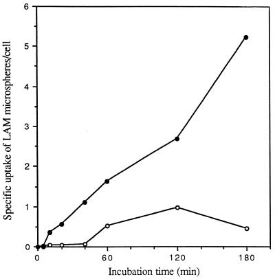
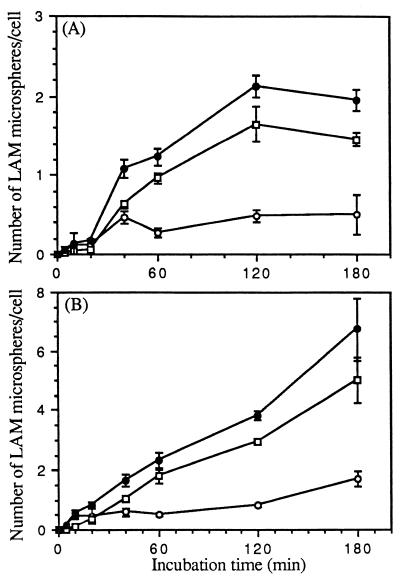
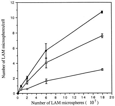
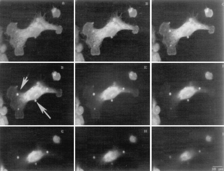
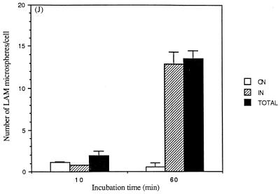
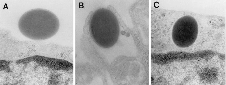
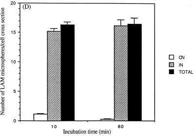
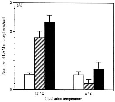
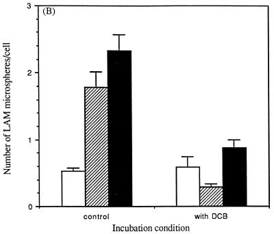
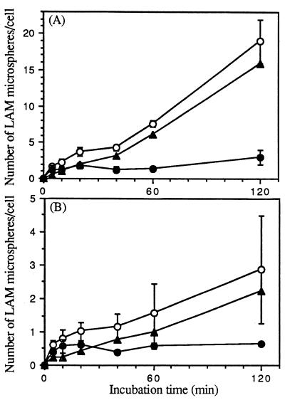
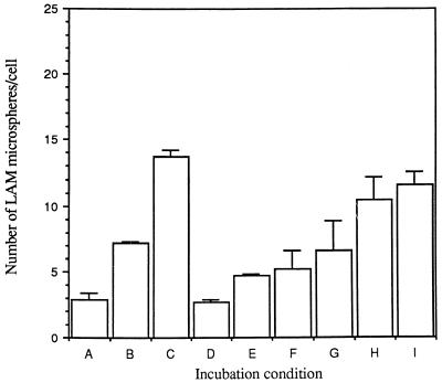
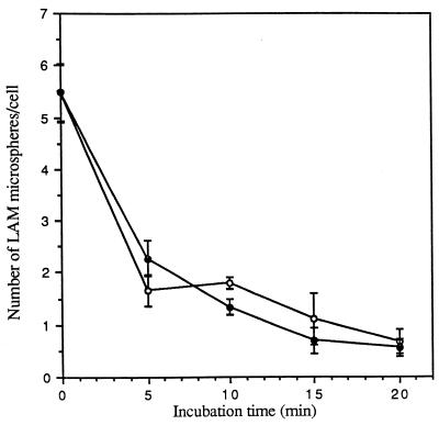
The macrophage mannose receptor (MR) along with complement receptors mediates phagocytosis of the M. tuberculosis virulent strains Erdman and H37Rv. We have determined that the terminal mannosyl units of the M. tuberculosis surface lipoglycan, lipoarabinomannan (LAM), from the Erdman strain serve as ligands for the MR. The biology of the MR (receptor binding and trafficking) in response to phagocytic stimuli is not well characterized. This study analyzes the MR-dependent phagocytosis mediated by Erdman LAM presented on a 1-micron-diameter phagocytic particle. Erdman LAM microspheres exhibited a time- and dose-dependent rapid increase in attachment and internalization by human monocyte-derived macrophages (MDMs). In contrast, internalization of LAM microspheres by monocytes was minimal. Microsphere internalization by MDMs was visualized and quantitated by immunofluorescence and confocal and electron microscopy and resembled conventional phagocytosis. Phagocytosis of LAM microspheres by MDMs was energy, cytoskeleton, and calcium dependent and was mannan inhibitable. Trypsin treatment of MDMs at 37 degrees C, which depleted surface and recycling intracellular pools of the MR, reduced the subsequent attachment of LAM microspheres. Trypsin treatment at 4 degrees C allowed for subsequent recovery of LAM microsphere phagocytosis at 37 degrees C by recycled MRs. Pretreatment of MDMs with cycloheximide influenced LAM microsphere phagocytosis to only a small extent, indicating that MR-dependent phagocytosis of the microspheres was occurring primarily by preformed recycled receptors. This study characterizes the requirements for macrophage phagocytosis of a LAM-coated particle mediated by the MR. This model will be useful in further characterization of the intracellular pathway taken by phagocytic particles coated with different LAM types in macrophages following ingestion.


Figures

FIG. 1
FIG. 1
Specificity of cell association of LAM microspheres with monocytes and MDMs. Monocytes and MDMs were incubated with 2 × 10⁷ LAM- or HSA (control)-coated microspheres for the indicated time at 37°C. At the end of each incubation period, monocytes and MDMs were fixed with formalin. The total number of LAM or HSA microspheres associated with the cell was enumerated by phase microscopy. The specific uptake of LAM microspheres by monocytes (open circles) and MDMs (solid circles) was obtained by subtracting the total number of associated HSA microspheres from that of LAM microspheres at each point for each cell type. All points are means from two independent experiments.
FIG. 2
FIG. 2
Time course of cell association of microspheres with monocytes and MDMs. Monocytes (A) and MDMs (B) were incubated with 2 × 10⁷ LAM microspheres for the indicated time at 37°C. At the end of each incubation period, monocytes and MDMs were washed and fixed with either 3.3% formalin (to visualize attached microspheres) or 100% methanol (to visualize all cell-associated microspheres). Microspheres associated with fixed monocytes and MDMs were visualized and enumerated by phase and indirect immunofluorescence microscopy with a primary polyclonal antibody against M. tuberculosis LAM and a fluorescein isothiocyanate-conjugated secondary antibody. The number of microspheres attached on the cell surface (open circles) or totally associated (solid circles) was measured. The number of internalized microspheres (open squares) was calculated. All points are means ± standard errors from two independent experiments.
FIG. 3
FIG. 3
Dose dependence of attachment and ingestion of LAM microspheres by MDMs. MDMs were incubated with the indicated number of LAM microspheres for 60 min at 37°C and then fixed with 3.3% formalin. Fixed MDMs were visualized as in Fig. 2, except that the number of total cell-associated microspheres was visualized and enumerated by phase microscopy instead of fluorescence microscopy. The number of microspheres attached to the cell (open circles) or totally associated (solid circles) was measured. The number of internalized microspheres (open squares) was calculated. All points are means ± standard errors from two independent experiments.
FIG. 4
FIG. 4
Confocal microscopy to examine internalization of LAM microspheres. Glass slides prepared for the experiments shown in Fig. 1 to 3 were examined by confocal microscopy. Serial sections of a single cell (A to I) were taken from the base of the cell to the apical surface. In panel D, the small arrow shows an internalized microsphere and the large arrow shows an attached microsphere (×1,000). Results are quantitated in panel J (for MDMs containing ≥1 microsphere). The number of microspheres attached (ON) and internalized (IN) and the total number of microspheres (TOTAL) were determined.
FIG. 4
FIG. 4
Confocal microscopy to examine internalization of LAM microspheres. Glass slides prepared for the experiments shown in Fig. 1 to 3 were examined by confocal microscopy. Serial sections of a single cell (A to I) were taken from the base of the cell to the apical surface. In panel D, the small arrow shows an internalized microsphere and the large arrow shows an attached microsphere (×1,000). Results are quantitated in panel J (for MDMs containing ≥1 microsphere). The number of microspheres attached (ON) and internalized (IN) and the total number of microspheres (TOTAL) were determined.
FIG. 5
FIG. 5
EM to examine internalization of LAM microspheres. MDMs on glass coverslips were incubated with LAM microspheres as in Fig. 1 for 60 min. MDMs were then fixed, prepared, and sectioned for analysis by transmission EM. (A) LAM microsphere attached to the cell surface (×40,000). (B) LAM microsphere in the process of phagocytosis (×40,000). (C) LAM microsphere within a phagosome (×40,000). Results are quantitated in panel D (for MDM cross sections containing ≥1 microsphere). The number of microspheres attached (ON) and internalized (IN) and the total number of microspheres (TOTAL) were determined.
FIG. 5
FIG. 5
EM to examine internalization of LAM microspheres. MDMs on glass coverslips were incubated with LAM microspheres as in Fig. 1 for 60 min. MDMs were then fixed, prepared, and sectioned for analysis by transmission EM. (A) LAM microsphere attached to the cell surface (×40,000). (B) LAM microsphere in the process of phagocytosis (×40,000). (C) LAM microsphere within a phagosome (×40,000). Results are quantitated in panel D (for MDM cross sections containing ≥1 microsphere). The number of microspheres attached (ON) and internalized (IN) and the total number of microspheres (TOTAL) were determined.
FIG. 6
FIG. 6
Temperature dependence and the role of the actin cytoskeleton in the cell association of LAM microspheres with MDMs. (A) MDMs were incubated with LAM microspheres for 60 min at 37 or 4°C. (B) MDMs were treated with dihydrocytochalasin B (5 μg/ml) for 30 min at 37°C and then incubated with LAM microspheres for 60 min at 37°C. At the end of each incubation period, MDMs were fixed with 3.3% formalin. Fixed MDMs were enumerated as described in the legend to Fig. 3. The number of microspheres attached to the cell (open bars) or totally associated (solid bars) was measured. The number of internalized microspheres (striped bars) was calculated. All points are means ± standard errors from two independent experiments.
FIG. 6
FIG. 6
Temperature dependence and the role of the actin cytoskeleton in the cell association of LAM microspheres with MDMs. (A) MDMs were incubated with LAM microspheres for 60 min at 37 or 4°C. (B) MDMs were treated with dihydrocytochalasin B (5 μg/ml) for 30 min at 37°C and then incubated with LAM microspheres for 60 min at 37°C. At the end of each incubation period, MDMs were fixed with 3.3% formalin. Fixed MDMs were enumerated as described in the legend to Fig. 3. The number of microspheres attached to the cell (open bars) or totally associated (solid bars) was measured. The number of internalized microspheres (striped bars) was calculated. All points are means ± standard errors from two independent experiments.
FIG. 7
FIG. 7
Time course of mannan-inhibitable cell association of LAM microspheres with MDMs. MDMs were incubated in the absence or presence of 4 mg of mannan per ml at 37 or 4°C and then incubated with 6 × 10⁷ microspheres for the indicated time at 37°C (A) or 4°C (B) and fixed in formalin. The number of total cell-associated microspheres was enumerated by phase-contrast microscopy. The specific uptake of microspheres (solid triangles) was obtained by subtracting the number of microspheres in the presence of mannan (solid circles) from that of microspheres in the absence of mannan (open circles). All points are means ± standard errors from two independent experiments.
FIG. 8
FIG. 8
Calcium dependence of cell association of LAM microspheres with MDMs. MDMs were incubated in calcium-free RPMI in the absence or presence of 1 mM EDTA with increasing concentrations of calcium for 30 min at 37°C. MDMs were then incubated with 6 × 10⁷ microspheres for 60 min at 37°C, washed, and fixed in formalin. The number of total cell-associated microspheres was enumerated by phase-contrast microscopy. (A) Ca2+-free medium. (B) Ca2+-free medium plus 1 mM Ca2+. (C) Ca2+-free medium plus 2 mM Ca2+. (D) EDTA (1 mM). (E) EDTA (1 mM) plus 0.125 mM Ca2+. (F) EDTA (1 mM) plus 0.25 mM Ca2+. (G) EDTA (1 mM) plus 0.5 mM Ca2+. (H) EDTA (1 mM) plus 1 mM Ca2+. (I) EDTA (1 mM) plus 2 mM Ca2+. All points are means ± standard errors from two independent experiments.
FIG. 9
FIG. 9
Effects of trypsin and EGTA on release of LAM microspheres attached to MDMs. MDMs were incubated with 6 × 10⁷ microspheres for 60 min at 4°C, washed, and then incubated in medium containing 0.01% trypsin (open circles) or 10 mM EGTA (solid circles) for the indicated time at 4°C, washed, and fixed in formalin. The number of total cell-associated microspheres was enumerated by phase-contrast microscopy. All points are means ± standard errors from two independent experiments.
FIG. 10
FIG. 10
Effect of treatment of MDMs with trypsin on the subsequent attachment of LAM microspheres. MDMs were incubated in the absence (control) or presence of 0.01% trypsin for the indicated times at 37°C and then incubated with 6 × 10⁷ microspheres for 20 min at 4°C, washed, and fixed in formalin. The condition chosen for trypsin treatment allowed the monolayer to remain intact. The number of total cell-associated microspheres was enumerated by phase-contrast microscopy, and the percentage of control was calculated for each time point (n = 2).
FIG. 11
FIG. 11
Removal of surface-expressed MR activity by trypsin treatment at 4°C and recovery of activity by warm up of the cell at 37°C. MDMs were incubated in the presence of 0.01% trypsin for the indicated time at 4°C (0 to 40 min, left side of the figure) and then incubated with 6 × 10⁷ microspheres for 60 min at 4°C and fixed in formalin, and the number of microspheres was enumerated. For the recovery assay (right side of the figure), MDMs were incubated in the presence of 0.01% trypsin for 40 min at 4°C, warmed for the indicated time (0 to 10 min) at 37°C, and then incubated with 6 × 10⁷ microspheres for 60 min at 4°C and fixed in formalin, and the number of microspheres was enumerated. All points are means ± standard errors from two independent experiments.

References

    1. Basu N, Sett R, Das P K. Down-regulation of mannose receptors on macrophages after infection with Leishmania donovani. Biochem J. 1991;277:451–456. - PMC - PubMed
    1. Brennan P J, Nikaido H. The envelope of mycobacteria. Annu Rev Biochem. 1995;64:29–63. - PubMed
    1. Chatterjee D, Lowell K, Rivoire B, McNeil M R, Brennan P J. Lipoarabinomannan of Mycobacterium tuberculosis. Capping with mannosyl residues in some strains. J Biol Chem. 1992;267:6234–6239. - PubMed
    1. Ezekowitz R A B, Sastry K, Bailly P, Warner A. Molecular characterization of the human macrophage mannose receptor: demonstration of multiple carbohydrate recognition-like domains and phagocytosis of yeasts in Cos-1 cells. J Exp Med. 1990;172:1785–1794. - PMC - PubMed
    1. Goren M B, Hart P D, Young M R, Armstrong J A. Prevention of phagosome-lysosome fusion in cultured macrophages by sulfatides of Mycobacterium tuberculosis. Proc Natl Acad Sci USA. 1976;73:2510–2514. - PMC - PubMed

Publication types

LinkOut - more resources