. 1998 Jun;66(6):2814-7.
doi: 10.1128/IAI.66.6.2814-2817.1998.

Listeria monocytogenes-infected hepatocytes are targets of major histocompatibility complex class Ib-restricted antilisterial cytotoxic T lymphocytes

Affiliations

Listeria monocytogenes-infected hepatocytes are targets of major histocompatibility complex class Ib-restricted antilisterial cytotoxic T lymphocytes

H G Bouwer et al. Infect Immun. 1998 Jun.

Abstract

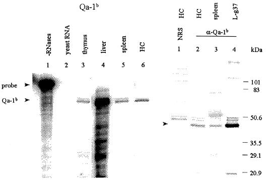
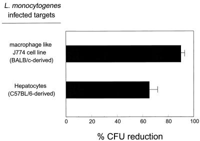
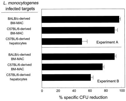
Subclinical infection of BALB/c mice with the intracellular bacterial pathogen Listeria monocytogenes results in the development of protective antilisterial immunity. L. monocytogenes can infect hepatocytes, and antilisterial cytotoxic T lymphocytes (CTL) lyse Listeria-infected hepatocytes in a major histocompatibility complex (MHC) class Ia-restricted manner. It remained to be determined whether L. monocytogenes-infected hepatocytes are susceptible to MHC class Ib-restricted cytolysis. In this study, we showed that hepatocytes express MHC class Ib molecule Qa-1(b) mRNA and protein. We further showed that Listeria-infected hepatocytes are susceptible to MHC class Ib-restricted cytolysis, since C57BL/6-derived Listeria-infected hepatocytes were lysed by BALB/c-derived antilisterial CTL. These results establish that Listeria-infected hepatocytes are susceptible to cytolysis by MHC class Ib restricted Listeria-specific CTL.


Figures

FIG. 1
FIG. 1
Hepatocytes express MHC class Ib molecules. (Left) Expression of Qa-1b mRNA as detected by RNase protection assay. Total RNA from the thymus (lane 3), liver (lane 4), spleen (lane 5), and hepatocytes (HC) (lane 6) was hybridized with a 360-nucleotide Qa-1b-specific probe. After RNase digestion, a 276-nucleotide Qa-1b-protected fragment was detected by using 6% denaturing acrylamide gels. RNase-treated (lane 2) and untreated (lane 1) yeast RNA-hybridized probes served as controls. The level of β-actin expression was used to measure the relative RNA content in each lane. (Right) Detection of the Qa-1b protein by immunoprecipitation. Freshly isolated hepatocytes (lanes 1 and 2), spleen cells (lane 3), and Qa-1b-transfected cell line L-g37 (lane 4) were metabolically labeled with 35S and lysed with Nonidet P-40 buffer. The lysates were precipitated with a rabbit anti-Qa-1b cytoplasmic tail serum (α-Qa-1b) (lanes 2 to 4). The band corresponding to Qa-1b is indicated by an arrowhead. Nonimmune rabbit serum (NRS) (lane 1) was used as a negative control.
FIG. 2
FIG. 2
Hepatocytes are susceptible to MHC class Ib-restricted cytolysis. Cells of the macrophage-like cell line J774 or hepatocytes from C57BL/6 mice were deposited at 105/well for use in a CFU reduction assay. These target cells were infected with L. monocytogenes at MOIs of 2 to 5 and 10 to 20, respectively. BALB/c-derived antilisterial CTL (E/T, 30:1) were added 3 h following infection; the assay was terminated 5 h later, and the remaining CFU were determined as described in Materials and Methods.
FIG. 3
FIG. 3
Hepatocytes are susceptible to L. monocytogenes-specific MHC class Ib-restricted cytolysis. BALB/c- or C57BL/6-derived BM-MAC or hepatocytes from C57BL/6 mice were obtained and deposited at 105/well for use in a CFU reduction assay. The target cells were infected with L. monocytogenes at an MOI of 10 to 20. BALB/c-derived nonimmune effector cells (E/T, 30:1) or L. monocytogenes-immune effector cells (E/T, 30:1) were added 3 h following infection; the assay was terminated 5 h later, and the remaining CFU were determined as described in Materials and Methods. Specific CFU reduction was determined by dividing the level of CFU reduction measured in target cell monolayers containing immune cells by the level of CFU reduction measured in target cell monolayers containing nonimmune cells. Nonspecific CFU reduction against Listeria-infected BM-MAC was less than 20% in both experiment A and experiment B. Nonspecific CFU reduction against Listeria-infected hepatocytes was 42% for experiment A and 37% for experiment B.

References

    1. Baldridge J R, Barry R A, Hinrichs D J. Expression of systemic protection and delayed-type hypersensitivity to Listeria monocytogenes is mediated by different T-cell subsets. Infect Immun. 1990;58:654–658. - PMC - PubMed
    1. Bishop D K, Hinrichs D J. Adoptive transfer of immunity to Listeria monocytogenes. The influence of in vitro stimulation on lymphocyte subset requirements. J Immunol. 1987;139:2005–2009. - PubMed
    1. Bouwer H G, Lindahl K F, Baldridge J R, Wagner C R, Barry R A, Hinrichs D J. An H2-T MHC class Ib molecule presents Listeria monocytogenes-derived antigen to immune CD8+ cytotoxic T cells. J Immunol. 1994;152:5352–5360. - PubMed
    1. Bouwer H G, Nelson C S, Gibbins B L, Portnoy D A, Hinrichs D J. Listeriolysin O is a target of the immune response to Listeria monocytogenes. J Exp Med. 1992;175:1467–1471. - PMC - PubMed
    1. Bouwer H G A, Barry R A, Hinrichs D J. Acquired immunity to an intracellular pathogen: immunologic recognition of L. monocytogenes-infected cells. Immunol Rev. 1997;158:137–146. - PubMed

Publication types