Comparative Study
. 1998 Jun;66(6):2845-53.
doi: 10.1128/IAI.66.6.2845-2853.1998.

Expression of two members of the pMGA gene family of Mycoplasma gallisepticum oscillates and is influenced by pMGA-specific antibodies

Affiliations
Comparative Study

Expression of two members of the pMGA gene family of Mycoplasma gallisepticum oscillates and is influenced by pMGA-specific antibodies

P F Markham et al. Infect Immun. 1998 Jun.

Abstract

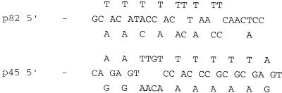
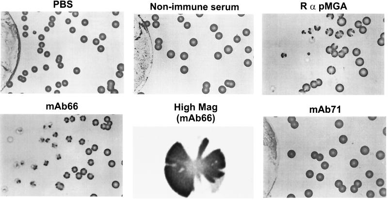
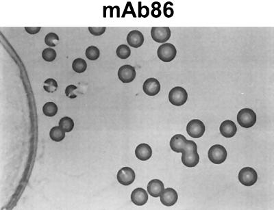
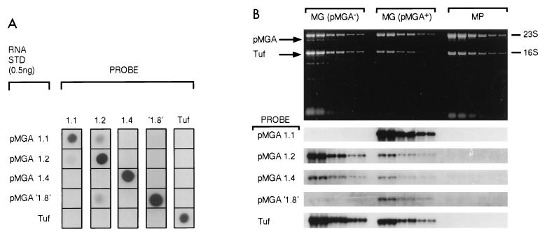
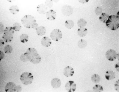
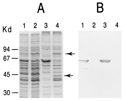
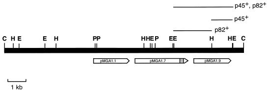
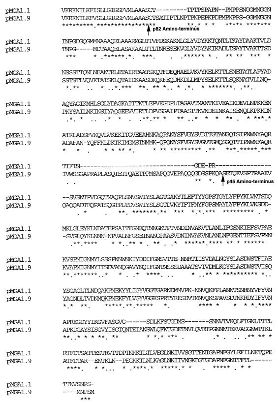
Certain monoclonal antibodies and polyclonal antisera directed to pMGA, the major protein of Mycoplasma gallisepticum, were tested for the ability to influence the surface phenotype of the cell population which resulted from their inclusion in growth medium. The polyclonal antiserum and one monoclonal antibody (MAb 66) resulted in an alteration of surface phenotype; specifically, populations of cells grown either on plates or in broth cultures which contained these reagents ceased the expression of pMGA and instead expressed an antigenically unrelated new polypeptide (p82). Upon the removal of antibody, the progeny of these cells regained pMGA expression and produced antigenically sectored colonies. The basis of this switch between pMGA+ and pMGA- states was shown to be transcriptional. The p82 polypeptide, the expression of which resulted from growth of cells in antibodies, was another member of the pMGA gene family and was located just downstream from the pMGA gene normally expressed by the M. gallisepticum cells used. Collectively the results of this work suggest that this organism has evolved an unusual means of altering the antigenic composition of its surface in response to antibodies or to other environmental cues.


Figures

FIG. 1
FIG. 1
Nucleotide sequences used for the oligonucleotide probes, which were based on amino acid sequences.
FIG. 2
FIG. 2
Nitrocellulose blots of colonies grown on agar culture plates around discs containing antibodies. Discs containing the reagents shown were placed at the centers of plates seeded with equal numbers of M. gallisepticum cells. After 6 days of growth, membrane lifts of each plate were immunostained with rabbit anti-pMGA (RαpMGA) as the primary detection reagent. In all cases, filter discs were located to the left of the colonies shown. The plate to the right of the MAb 66 panel depicts a single colony from this plate at a 10-fold-higher magnification.
FIG. 2
FIG. 2
Nitrocellulose blots of colonies grown on agar culture plates around discs containing antibodies. Discs containing the reagents shown were placed at the centers of plates seeded with equal numbers of M. gallisepticum cells. After 6 days of growth, membrane lifts of each plate were immunostained with rabbit anti-pMGA (RαpMGA) as the primary detection reagent. In all cases, filter discs were located to the left of the colonies shown. The plate to the right of the MAb 66 panel depicts a single colony from this plate at a 10-fold-higher magnification.
FIG. 3
FIG. 3
SDS-PAGE and Western blot analysis of M. gallisepticum cultured in the presence of a pMGA-specific MAb. (a) Coomassie blue-stained molecular weight standards (lane 1) and cells grown in broth medium alone (lane 2) or grown in broth containing MAb 66 (lane 3). (b) Rabbit anti-pMGA serum was used to immunostain a Western transfer of cellular proteins from mycoplasma cells grown in broth alone (lane 1) or in broth containing MAb 66 (lane 2). Arrowheads indicate the location of the pMGA protein. Arrows indicate the positions of 45- and 82-kDa bands referred to in the text. Molecular weight protein standards (Bio-Rad) are phosphorylase b (97,400), BSA (66,200), ovalbumin (45,000), and carbonic anhydrase (31,000).
FIG. 4
FIG. 4
Growth of M. gallisepticum cells in MAb 66-containing medium results in a selective cessation of transcription of the pMGA1.1 gene. (A) Synthetic sense-strand RNA samples (0.5 ng) derived from the four pMGA genes indicated and from the elongation factor Tuf gene were blotted (horizontally) onto nylon membrane strips. Each strip was hybridized with the indicated labeled probes (vertically). Details of synthetic RNA molecules, probes, and hybridization conditions are from reference . STD, standard. (B) Twofold serial dilutions of purified RNA from M. gallisepticum cells grown in medium to which MAb 66 had been added (5 μg ml−1) [MG (pMGA−1)] or in the medium without additive [MG (pMGA+)] or from Mycoplasma pullorum cells (MP) were subjected to denaturing electrophoresis, and replicate gels were either photographed to detect ethidium bromide patterns (upper panel) or subjected to Northern blot analysis using the labeled probes indicated alongside strips on the lower panel. Hybridization conditions used for individual probes were identical to those in panel A. Binding of probes was detected and then quantified with a phosphorimager.
FIG. 5
FIG. 5
Immunostaining of nitrocellulose blots of M. gallisepticum colonies derived from pMGA cells. The expression of pMGA in colonies derived from cells originally grown in broth containing MAb 66 was detected by immunostaining nitrocellulose blots of colonies with rabbit anti-pMGA serum. Sectorial reacquisition of pMGA expression is apparent.
FIG. 6
FIG. 6
The p82 and p45 polypeptides are partitioned into Triton X-114. M. gallisepticum cells were grown either in broth medium containing no antibody additive (lanes 1 and 3) or in medium containing 5 μg of MAb 66 ml−1 (lanes 2 and 4). Recovered cells were boiled in SDS-containing buffer and subjected to SDS-PAGE (lanes 1 and 2). Alternatively, cells were lysed with Triton X-114 and subjected to partition into detergent-rich phases before electrophoresis (lanes 3 and 4). (A) Coomassie blue-stained gel; (B) identical gel transferred to a PVDF membrane and then immunostained with rabbit anti-pMGA followed by HRPO-conjugated anti-rabbit Ig. Arrowheads indicate the p45 and p82 species referred to in the text.
FIG. 7
FIG. 7
Restriction map of a ClaI fragment containing pMGA1.1, pMGA1.7, p82, and p45 sequences. Single and double digests of the cloned 13-kb ClaI fragment containing the pMGA1.1 coding sequence were conducted with the restriction enzymes ClaI (C), EcoRI (E), HindIII (H), and PstI (P). Digests were subjected to agarose gel electrophoresis for fragment size measurements, and the resultant gels were subjected to Southern blotting. The p82 and p45 N-terminal oligonucleotide probes used in Southern blot studies were based on the amino acid sequences in Table 1 and are listed in Fig. 1. Informative fragments which bound the p82 and p45 probes are indicated. The approximate lengths and locations of the three pMGA gene coding sequences referred to in the text are shown. The shaded bar toward the 3′ end of the pMGA1.7 gene depicts an unusual DNA insertion which has not yet been definitively sequenced.
FIG. 8
FIG. 8
Predicted amino acid sequences of the pMGA1.9 polypeptide and its homology to pMGA1.1. The predicted amino acid sequence of pMGA1.9 is aligned to the known sequence of pMGA1.1 to maximize homology. Residues identical with counterparts in pMGA1.1 are marked with asterisks and conservative substitutions are indicated with dots. The predicted amino termini of the p82 and p45 polypeptides are indicated by arrows.
FIG. 9
FIG. 9
Sequence alignment between 5′ noncoding regions of pMGA genes. The nucleotide sequences compared extend from the stop codons of preceding coding sequences and extend to the translational start codons of the pMGA genes compared. The −35 and −10 regions of putative promoters are boxed, and the GAA repeat segments of each region occur 5′ to the −35 regions.

References

    1. Bassegio N B, Glew M D, Markham P F, Whithear K G, Browning G F. Size and genomic location of the pMGA multigene family of Mycoplasma gallisepticum. Microbiology. 1995;142:1429–1435. - PubMed
    1. Bhugra B, Voelker L L, Zou N, Yu H, Dybwig K. Mechanism of antigenic variation in Mycoplasma pulmonis: interwoven site-specific DNA inversions. Mol Microbiol. 1995;18:703–714. - PubMed
    1. Bordier C. Phase separation of integral membrane proteins in Triton X-114 solution. J Biol Chem. 1981;256:1604–1607. - PubMed
    1. Chomczynski P, Sacchi N. Single step method of RNA isolation by acid guanidinium thiocyanate-phenol-chloroform extraction. Anal Biochem. 1987;162:156–159. - PubMed
    1. Church G M, Gilbert W. Genomic sequencing. Proc Natl Acad Sci USA. 1984;81:1991–1995. - PMC - PubMed

Publication types

MeSH terms

Associated data

LinkOut - more resources