. 1998 Jun;66(6):2895-904.
doi: 10.1128/IAI.66.6.2895-2904.1998.

Characterization of protective epitopes in a highly conserved Plasmodium falciparum antigenic protein containing repeats of acidic and basic residues

Affiliations

Characterization of protective epitopes in a highly conserved Plasmodium falciparum antigenic protein containing repeats of acidic and basic residues

P Sharma et al. Infect Immun. 1998 Jun.

Abstract

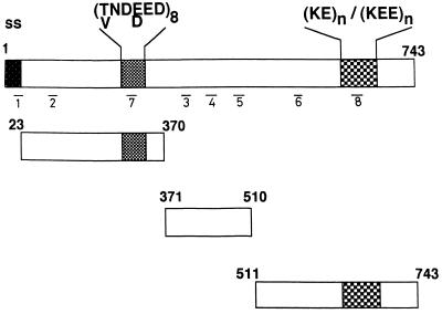
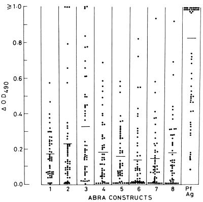
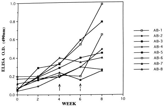
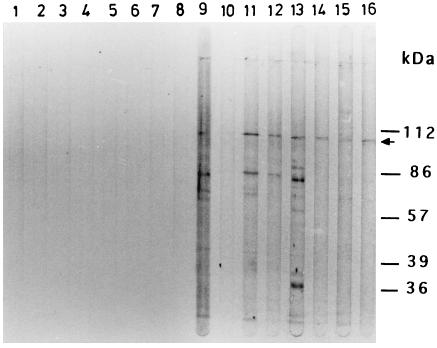
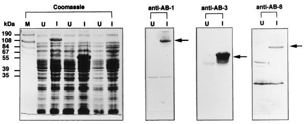
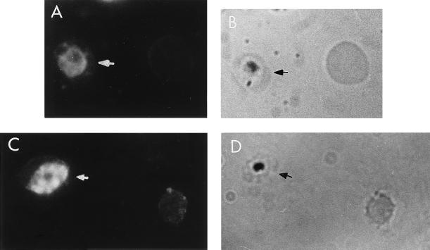
The delineation of putatively protective and immunogenic epitopes in vaccine candidate proteins constitutes a major research effort towards the development of an effective malaria vaccine. By virtue of its role in the formation of the immune clusters of merozoites, its location on the surface of merozoites, and its highly conserved nature both at the nucleotide sequence level and the amino acid sequence level, the antigen which contains repeats of acidic and basic residues (ABRA) of the human malaria parasite Plasmodium falciparum represents such an antigen. Based upon the predicted amino acid sequence of ABRA, we synthesized eight peptides, with six of these (AB-1 to AB-6) ranging from 12 to 18 residues covering the most hydrophilic regions of the protein, and two more peptides (AB-7 and AB-8) representing its repetitive sequences. We found that all eight constructs bound an appreciable amount of antibody in sera from a large proportion of P. falciparum malaria patients; two of these peptides (AB-1 and AB-3) also elicited a strong proliferation response in peripheral blood mononuclear cells from all 11 human subjects recovering from malaria. When used as carrier-free immunogens, six peptides induced a strong, boostable, immunoglobulin G-type antibody response in rabbits, indicating the presence of both B-cell determinants and T-helper-cell epitopes in these six constructs. These antibodies specifically cross-reacted with the parasite protein(s) in an immunoblot and in an immunofluorescence assay. In another immunoblot, rabbit antipeptide sera also recognized recombinant fragments of ABRA expressed in bacteria. More significantly, rabbit antibodies against two constructs (AB-1 and AB-5) inhibited the merozoite reinvasion of human erythrocytes in vitro up to approximately 90%. These results favor further studies so as to determine possible inclusion of these two constructs in a multicomponent subunit vaccine against asexual blood stages of P. falciparum.


Figures

FIG. 1
FIG. 1
Schematic representation of P. falciparum ABRA and its three fragments obtained by PCR amplification of the parasite genomic DNA and expressed as recombinant proteins as described in Materials and Methods. The small, numbered horizontal bars indicate the positions of the sequences chosen for synthetic peptides AB-1 to AB-8 as described in Materials and Methods. ss, signal sequence.
FIG. 2
FIG. 2
Distribution profile of antibody levels obtained by ELISA using sera from 50 clinical malaria patients. Each serum was diluted 1/200 and tested in duplicate against each of the eight ABRA constructs. The ΔOD490 value was obtained by subtracting the OD490 value given by a normal human serum pool from that given by the respective clinical serum. Each data point represents a mean of duplicate values. A ΔOD490 value of 0.2 or more was defined as positive. The solid horizontal line in each column represents the mean ΔOD490 value. Pf Ag, P. falciparum antigen.
FIG. 3
FIG. 3
Time course of IgG-type antibody responses generated in rabbits as monitored by an ELISA using homologous peptides as the capturing antigens. Rabbits immunized with the carrier-free ABRA peptides received boosters at week 4 (solid arrow); animals immunized with AB-2, AB-6, and AB-7 received boosters one more time at week 6 (broken arrow). Each serum was tested at a single dilution of 1/200 in duplicate wells.
FIG. 4
FIG. 4
Cross-reactivity of rabbit antipeptide antibodies with the parasite protein(s) as monitored in an ELISA using parasite lysate prepared from the asexual blood stages of P. falciparum as the capturing antigen. Each serum was tested at a single dilution of 1/200 in duplicate wells. See the legend to Fig. 3 for additional details.
FIG. 5
FIG. 5
Cross-reactivity of rabbit antipeptide sera with the parasite protein(s) in an immunoblot assay. The parasite proteins extracted from the asexual blood stages of P. falciparum were separated on an SDS–10% PAGE gel, transferred onto a nitrocellulose membrane, and probed with different antipeptide sera by using a Bio-Rad Mini Protean II Multi-Screen apparatus. Lanes 1 to 8 were probed with the preimmune sera, and lanes 9 to 16 were probed with the respective test sera. Thus, the lanes are for sera as follows: 1 and 9, AB-1 sera; 2 and 10, AB-2 sera; 3 and 11, AB-3 sera; 4 and 12, AB-4 sera; 5 and 13, AB-5 sera; 6 and 14, AB-6 sera; 7 and 15, AB-7 sera; and 8 and 16, AB-8 sera. Apart from the specific protein at approximately 101 kDa (arrow), some other bands were also detected, which may be degradation products of ABRA.
FIG. 6
FIG. 6
Three recombinant constructs representing the N-terminal region (AB-N), the middle region (AB-M), and the C-terminal region (AB-C) of ABRA were expressed as recombinant fusion proteins in E. coli under conditions of IPTG induction (lanes I) and immunoblotted with rabbit anti-AB-1 (AB-1), anti-AB-3 (AB-3), and anti-AB-8 (AB-8) sera. ABRA constructs of the expected sizes (arrows) were specifically recognized by the respective sera; no such reactivity was noticed in the control, uninduced bacterial cultures (lanes U).
FIG. 7
FIG. 7
Immunofluorescence on air-dried, acetone-fixed monolayers of P. falciparum-infected erythrocytes (arrows) probed with rabbit anti-AB-1 serum. Shown are a brightly fluorescent trophozoite (A and B) and merozoites (C and D) within a mature schizont seen under UV illumination (A and C) and visible light (B and D); uninfected erythrocytes which did not react with antibody are also seen.

References

    1. Alonso P L, Smith T, Armstrong Schellenberg J R M, Masanja H, Mwankusye S, Urassa H, Bastos de Azevedo I, Chongela J, Kobero S, Menendez C, Hurt N, Thomas M C, Lyimo E, Weiss N A, Hayes R, Kitua A Y, Lopez M C, Kilama W L, Teuscher T, Tanner M. Randomised trial of efficacy of SPf66 vaccine against Plasmodium falciparum malaria in children in southern Tanzania. Lancet. 1994;344:1175–1181. - PubMed
    1. Anders R F. Multiple cross-reactivities amongst antigens of Plasmodium falciparum impair the development of protective immunity against malaria. Parasite Immunol. 1986;8:529–539. - PubMed
    1. Anders R F, Smythe J A. Polymorphic antigens in Plasmodium falciparum. Blood. 1989;74:1865–1875. - PubMed
    1. Banyal H S, Misra G C, Gupta C M, Dutta G P. Involvement of malaria proteases in the interaction between the parasite and the host erythrocyte in Plasmodium knowlesi infections. J Parasitol. 1980;67:623–626. - PubMed
    1. Biggs B A, Gooze L, Wycherly K, Wollish W, Southwell B, Leech J H, Brown G V. Antigenic variation in Plasmodium falciparum. Proc Natl Acad Sci USA. 1991;88:9171–9174. - PMC - PubMed

Publication types

LinkOut - more resources