. 1998 Jun;66(6):2922-7.
doi: 10.1128/IAI.66.6.2922-2927.1998.

In vivo binding of immunoglobulin M to the surfaces of Babesia bigemina-infected erythrocytes

Affiliations

In vivo binding of immunoglobulin M to the surfaces of Babesia bigemina-infected erythrocytes

I E Echaide et al. Infect Immun. 1998 Jun.

Abstract

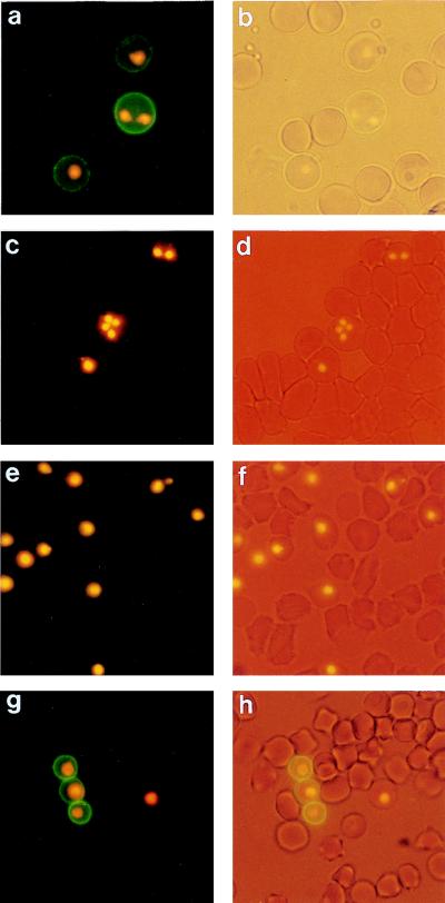
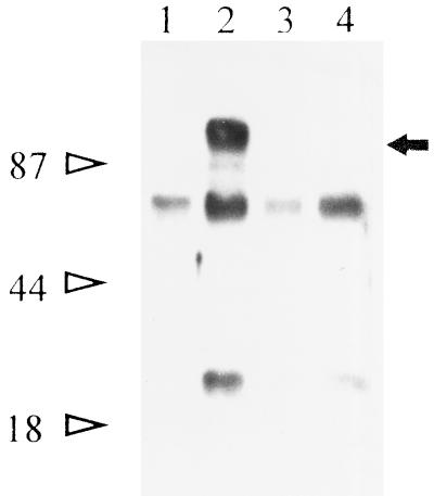
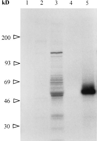
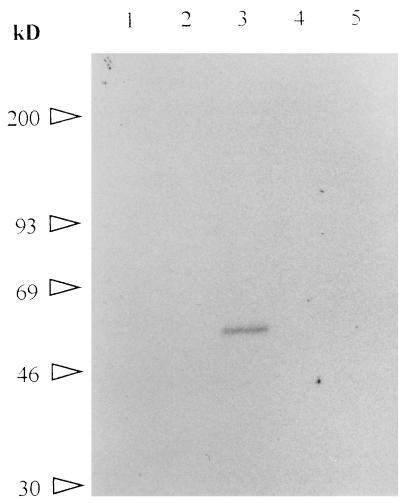
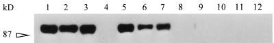
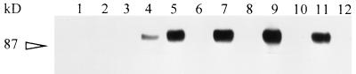
Babesia bigemina infection of mature bovine erythrocytes results in new proteins specifically exposed on the parasitized cell surface. Monoclonal antibody (MAb) 64/32 binds a protein, designated p94, on B. bigemina-infected erythrocytes but not on either uninfected or B. bovis-parasitized erythrocytes. However, p94 was not encoded by B. bigemina and was not a parasite-modified erythrocyte membrane protein. In contrast, we showed that p94 could be eluted from the infected erythrocyte surface and was identified as specifically bound immunoglobulin M (IgM) heavy chain for the following reasons: (i) MAb 64/32 bound a reduced molecule of 94 kDa in both infected erythrocyte lysates and normal bovine serum; (ii) MAb 64/32 bound a 94-kDa molecule in reduced preparations of purified IgM; (iii) an anti-bovine mu heavy-chain MAb, BIg73, reacted specifically with the surface of infected erythrocytes and bound the 94-kDa molecule in lysates of infected erythrocytes, normal bovine serum, and purified IgM; and (iv) immunoprecipitation of infected erythrocyte lysates with MAb 64/32 depleted the 94-kDa antigen bound by anti-mu MAb BIg73 and vice versa. Binding of IgM to the infected erythrocyte surface was detected in vivo early in acute parasitemia and occurred during both the trophozoite and merozoite stages of intraerythrocytic parasitism. The common feature of IgM binding to the parasitized erythrocyte surface among otherwise genetically and antigenically distinct B. bigemina strains is suggestive of an advantageous role in parasite survival in vivo.


Figures

FIG. 1
FIG. 1
Reactivity of MAbs with the surfaces of B. bigemina-infected erythrocytes assessed by live-cell IFA. (a to f) In vitro culture. (a) IFA reactivity of MAb 64/32; (b) light microscopy of the same field; (c) lack of reactivity of MAb 64/32 on the surfaces of B. bovis-infected erythrocytes; (d) light microscopy of the same field as in panel c; (e) lack of reactivity of the isotype control MAb Tryp1E1; (f) light microscopy of the same field as in panel e. (g and h) In vivo infection with B. bigemina (day 3). (g) IFA reactivity of MAb 64/32; (h) light microscopy of the same field as in panel g. Parasite nuclei were stained with ethidium bromide to detect infected erythrocytes.
FIG. 2
FIG. 2
Immunoprecipitation of antigens from the surfaces of B. bigemina-infected erythrocytes. Biotin-labeled surface proteins from infected erythrocytes were immunoprecipitated with MAb 64/32 (lane 2, arrow) or the negative control MAb 64/11 (lane 4). Surface-labeled uninfected erythrocytes were immunoprecipitated with either MAb 64/32 (lane 1) or MAb 64/11 (lane 3). Molecular size standards are designated on the left.
FIG. 3
FIG. 3
Immunoprecipitation of 35S-labeled B. bigemina proteins. Proteins labeled with [35S]methionine during in vitro growth of B. bigemina were immunoprecipitated with MAb 64/32 (lane 4), MAb 14/16 (anti-p58, RAP-1) (lane 5), murine polyclonal antiserum to p94 (lane 2), bovine postinfection serum B240 (lane 3), or preinoculation murine serum (lane 1). Molecular size standards are designated on the left.
FIG. 4
FIG. 4
Immunoprecipitation of 3H-labeled B. bigemina proteins. Proteins labeled with a mixture of 15 tritiated amino acids during in vitro growth of B. bigemina were immunoprecipitated with MAb 64/32 (lane 1), MAb 14/16 (lane 3), murine polyclonal antiserum to p94 (lane 5), isotype control MAb Tryp1E1 (lane 2), or preinoculation murine serum (lane 4). Molecular size standards are designated on the left.
FIG. 5
FIG. 5
Reactivity of MAb 64/32 with infected erythrocytes, normal bovine serum, and purified IgM. B. bigemina-infected erythrocytes (lanes 1, 5, and 9), normal bovine serum (lanes 2, 6, and 10), purified bovine IgM (lanes 3, 7, and 11), or purified bovine IgG (lanes 4, 8, and 12) were separated by SDS-PAGE under reducing conditions. The immunoblotted proteins were probed with MAb 64/32 (lanes 1 to 4), MAb BIg73 (anti-bovine μ heavy chain) (lanes 5 to 8), or isotype control MAb Tryp1E1 (lanes 9 to 12). The 87-kDa molecular size standard is indicated on the left.
FIG. 6
FIG. 6
Binding of IgM from B. bigemina-infected erythrocytes by MAbs 64/32 and BIg73. Biotin-labeled infected erythrocyte lysates were specifically depleted by immunoprecipitation with the first MAb; the proteins remaining in the supernatant were then immunoprecipitated by the second MAb. The following six pairs of MAbs were used in a “first-second” order for the immunoprecipitations: for uninfected erythrocytes, ANA8-64/32 (lanes 1 and 2); for infected erythrocytes; 64/11-64/32 (lanes 3 and 4), 64/32-64/11 (lanes 5 and 6), BIg73-BIg73 (lanes 7 and 8), BIg73-64/32 (lanes 9 and 10), and 64/32-BIg73 (lanes 11 and 12). BIg73 is against the bovine μ heavy chain; MAbs ANA8 and 64/11 are isotype controls. The 87-kDa molecular size standard is indicated on the left.

Similar articles

Cited by

References

    1. Adams J H, Hudson D E, Torii M, Ward G E, Wellems T E, Aikawa M, Miller L H. The Duffy receptor family of Plasmodium knowlesi is located within the micronemes of invasive malaria merozoites. Cell. 1990;63:141–153. - PubMed
    1. Aikawa M, Rabbege J, Uni S, Ristic M, Miller L H. Structural alteration of the membrane of the erythrocytes infected with Babesia bovis. Am J Trop Med Hyg. 1985;34:45–49. - PubMed
    1. Baruch D, Pasloske B L, Singh H B, Bi X, Ma X C, Feldman M, Taraschi T F, Howard R J. Cloning the P. falciparum gene encoding PfEMP1, a malarial variant antigen and adherence receptor on the surface of parasitized human erythrocytes. Cell. 1995;82:77–87. - PubMed
    1. Berendt A R, Ferguson D J P, Gardner J, Turner G, Rowe A, McCormick C, Roberts D, Craig A, Pinches R, Elford B C, Newbold C I. Molecular mechanisms of sequestration in malaria. Parasitol. 1990;108:S19–S28. - PubMed
    1. Crandall I, Guthrie N, Sherman I W. Plasmodium falciparum: sera of individuals living in a malaria-endemic region recognize peptide motifs of the human erythrocyte anion transport protein. Am J Trop Med Hyg. 1995;52:450–452. - PubMed

Publication types

MeSH terms