. 1998 Jun;66(6):2943-50.
doi: 10.1128/IAI.66.6.2943-2950.1998.

Helicobacter pylori disrupts epithelial barrier function in a process inhibited by protein kinase C activators

Affiliations

Helicobacter pylori disrupts epithelial barrier function in a process inhibited by protein kinase C activators

A M Terrés et al. Infect Immun. 1998 Jun.

Abstract

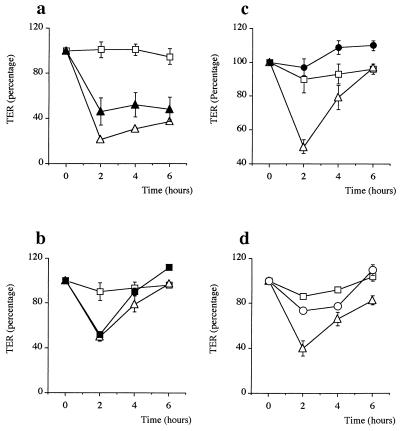
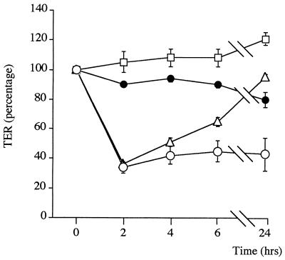
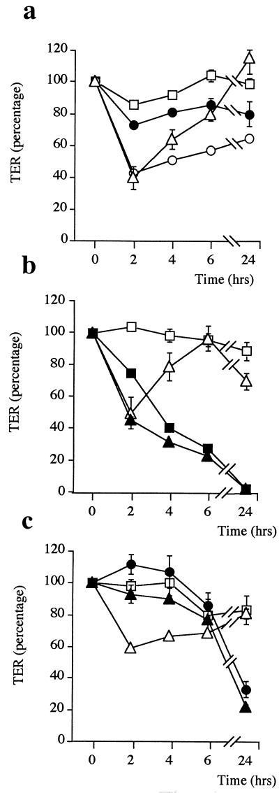
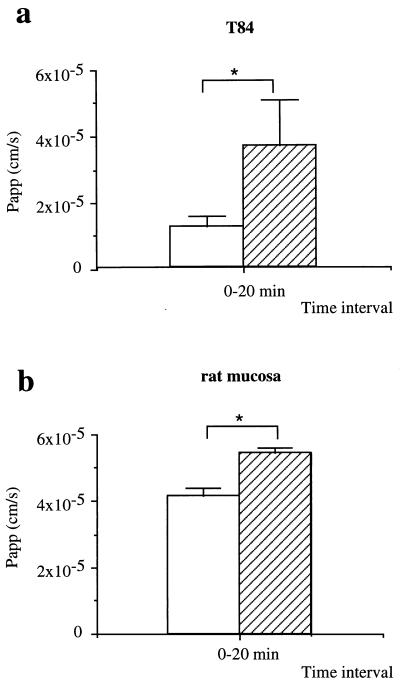
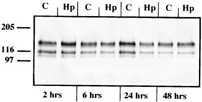
Helicobacter pylori colonizes the gastric mucosa, and the infection is related to the development of diverse gastric pathologies, possibly by directly or indirectly affecting epithelial-cell function. We analyzed the influence of the bacteria on transepithelial electrical resistance (TER) on a model tight epithelium, T84, grown to confluence in permeable filters. H. pylori sonicates produced a dramatic decrease in TER after 1 to 2 h of exposure, while sonicates from other bacteria did not induce a significant reduction of TER. The effect induced by sonicates was mimicked by a water-soluble fraction from the bacterial surface, was not reproducible with isolated lipopolysaccharide, and was concomitant with a significant increase in the paracellular permeability of the marker molecule [14C]mannitol. Furthermore, H. pylori sonicates also provoked a significant increase in permeability to [14C]mannitol across rat gastric mucosa in vitro. The sonicate-induced decrease in TER in T84 monolayers was inhibited by the protein kinase C (PKC) activator phorbol myristate acetate. As PKC is directly involved in tight junction regulation, we suggest that H. pylori may induce intracellular signalling events counteracting PKC effects. Following long-term H. pylori stimulation, epithelial monolayers regained baseline resistance values slowly after 24 h. The resistance recovery process was inhibited by cycloheximide, indicating its dependency upon protein synthesis. No association between resistance variation and E-cadherin protein levels was observed. These results indicate that H. pylori alters in vitro the barrier properties of the epithelium, probably by generating cell signalling events counteracting the normal function of PKC. This increased permeability may provide a potential mechanism by which H. pylori antigens can reach the gastric lamina propria, thereby activating the mucosal immune system.


Figures

FIG. 1
FIG. 1
TER variation at different time intervals. TER was monitored in T84 monolayers treated with 5 (open circles), 10 (solid triangles), and 15 (solid squares) μg of sonicated NCTC 11638 H. pylori/ml versus control cells in medium alone (open squares). The effect is concentration dependent, and the maximum decrease in TER occurred between 1 and 2 h in monolayers treated with 15 μg of H. pylori/ml. The graph shown is from a representative experiment; P = 0.008; n = 3). (Inset) TER of control monolayers (open bar) and of monolayers basolaterally treated with H. pylori (striped bar). No significant difference was found.
FIG. 2
FIG. 2
TER variation of T84 monolayers exposed to 15 μg of E. coli (solid circles), C. jejuni (open triangles), or H. influenzae (solid square) sonicate/ml compared to cells in medium alone (open squares) and cells treated with H. pylori NCTC 11638 sonicate (open circles). H. pylori produced the most significative TER at 2 h. Data from a representative experiment are shown (n = 6).
FIG. 3
FIG. 3
Variation of TER of T84 monolayers exposed to whole NCTC 11638 bacteria (solid triangles) (a), H. pylori NCTC 11638 surface proteins (solid squares) (b), H. pylori LPS (solid circles) (c), or heat-inactivated H. pylori NCTC 11638 sonicate (open circles) (d) compared to TERs of control cells in medium alone (open squares) and cells exposed to NCTC 11638 sonicate (open triangles). Whole bacteria and surface proteins mimicked the effect of H. pylori sonicate, while LPS had no effect. When H. pylori sonicate is heated at 70°C for 15 min, the characteristic TER drop at 2 h does not occur, which indicates that the molecule(s) involved in this process is proteinic in nature. Data shown are from four representative experiments.
FIG. 4
FIG. 4
Variation of TER in T84 monolayers stimulated with 15 μg of sonicated H. pylori NCTC 11638/ml in the presence of the protein synthesis inhibitor cycloheximide (open circles) versus T84 monolayers with cycloheximide alone (solid circles), medium alone (open squares), and H. pylori sonicates alone (open triangles). Cycloheximide did not prevent the decrease in TER produced by H. pylori sonicate at 2 h but did block the subsequent reestablishment of the pretreatment TER baseline, indicating that the process is dependent upon protein synthesis. Data shown are from a representative experiment (n = 3).
FIG. 5
FIG. 5
Variation of TER in T84 monolayers stimulated with H. pylori sonicate plus the protein tyrosine kinase inhibitor herbimycin A (open circles) versus herbimycin alone (solid circles) (a), the PKC inhibitor staurosporine (solid triangles) versus staurosporine alone (solid squares) (b), or the PKC activator PMA (solid triangles) versus PMA alone (solid circles) (c). Open squares, TER of control cells in medium alone; open triangles, TER of cells in the presence of H. pylori sonicate alone. Herbimycin A did not prevent the decrease in TER produced by H. pylori sonicate at 2 h, but it affected the subsequent reestablishment of TER. Staurosporine by itself produced the abolishment of the resistance at 24 h. In contrast, in the presence of PMA, the characteristic early drop in TER produced by H. pylori sonicate did not occur. Data shown are from four representative experiments (n = 3 to 6).
FIG. 6
FIG. 6
H. pylori-induced increase in permeability. (a) T84 confluent monolayers. The Papp of [14C]mannitol flux in non-H. pylori-treated control cells (open bar) versus that for H. pylori-pretreated cells (striped bar) is shown. (b) Rat mucosa. The Papp of [14C]mannitol flux in control nontreated rat mucosa (open bar) versus that for H. pylori-pretreated rat mucosa (striped bar) is shown. In paired experiments, basolateral-to-apical [14C]mannitol flux measurements were made over a single 20-min period following 2-h exposure of T84 cells or rat mucosa to H. pylori sonicates (15 μg/ml). Graphs show Papp means from six (a) and four (b) experiments. P < 0.05 for panel a and P < 0.05 for panel b by the Mann-Whitney U test.
FIG. 7
FIG. 7
Response of T84 monolayers to established secretagogues. Control (open circles) and H. pylori-pretreated (solid circles) confluent T84 monolayers mounted on Ussing chambers were stimulated with 3 and 10 μM forskolin and 10 and 100 μM carbachol at the indicated times. Treated and control monolayers were able to respond to both secretagogues, and the highest stimulation was obtained with 10 μM carbachol. Changes in SCC are indicative of the relative abilities of the monolayers to secrete chloride ions.
FIG. 8
FIG. 8
Western blot analysis of E-cadherin expression in T84 cells treated with H. pylori (Hp) for 2, 6, 24, and 48 h and in control cells (C) in medium alone. Besides the classical 120-kDa band, two smaller bands (∼100 and ∼105 kDa) were detected in all cases. Western blot analysis detected no change in the basal levels of E-cadherin after H. pylori treatment at any of the times at which they were examined.
FIG. 9
FIG. 9
Immunoprecipitation-Western blot analysis of tyrosine phosphorylation of E-cadherin in control and H. pylori-treated T84 monolayers. Western blots were probed with anti-E-cadherin antibody (a) and antiphosphotyrosine antibody (b). Lanes: A, control T84 whole-cell preparation; B, control T84 cells immunoprecipitated with anti-E-cadherin MAb; C, H. pylori-treated T84 cells immunoprecipitated with anti-E-cadherin MAb. E-cadherin (120 kDa) was not tyrosine phosphorylated in any case, but tyrosine phosphorylation was detected on two coprecipitated proteins (94 kDa, likely to be β-catenin, and 147 kDa). Phosphorylation of these proteins was not altered by exposure to H. pylori.
FIG. 10
FIG. 10
Anti-E-cadherin immunofluorescence staining of T84 monolayers grown in plastic chamber slides of cells treated with H. pylori sonicates for 2 h (A) versus control cells in medium alone (B). Treated cells appeared bigger than controls at 2 h. Changes in morphology and loss of adherence to plastic are maximum at 48 h. Bar, 40 μm.

References

    1. Aghib D, McCrea P. The E-cadherin complex contains the src substrate p120. Exp Cell Res. 1995;218:359–369. - PubMed
    1. Anderson J, Itallie C V. Tight junctions and the molecular basis for regulation of paracellular permeability. Am J Physiol. 1995;269:G467–G475. - PubMed
    1. Balda M, Gonzales-Mariscal L, Contreras R, Macias-Silva M, Torres-Markez M, Garcia-Sainz J, Cereijido M. Assembly and sealing of tight junctions: possible participation of G-proteins, phospholipase C, protein kinase C and calmodulin. J Membr Biol. 1991;122:193–202. - PubMed
    1. Balda M, Gonzalez M, Matter K, Cereijido M, Anderson J. Assembly of the tight junction: the role of diacylglycerol. J Cell Biol. 1993;123:293–302. - PMC - PubMed
    1. Borenfreund E, Puerner J. A simple quantitative procedure using monolayer cultures for cytotoxicity assays (9HTD/NR-90) J Tissue Cult Methods. 1984;9:7–9.

Publication types