. 1998 Jun;66(6):2951-9.
doi: 10.1128/IAI.66.6.2951-2959.1998.

Evaluation of new vaccines in the mouse and guinea pig model of tuberculosis

Affiliations

Evaluation of new vaccines in the mouse and guinea pig model of tuberculosis

S L Baldwin et al. Infect Immun. 1998 Jun.

Abstract

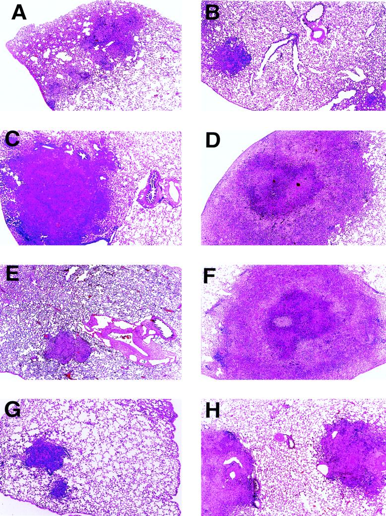
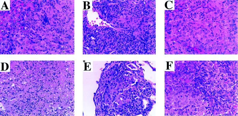
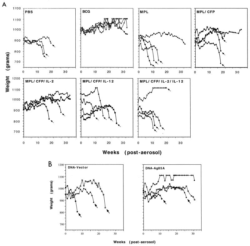
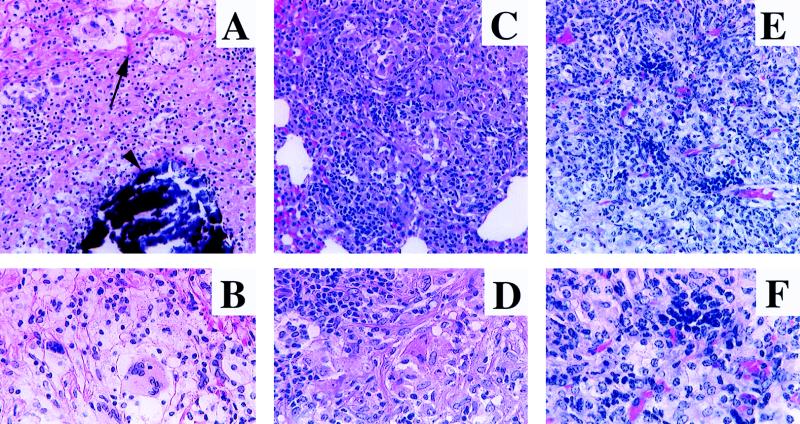
The results of this study provide the first evidence that two completely separate vaccine approaches, one based on a subunit vaccine consisting of a mild adjuvant admixed with purified culture filtrate proteins and enhanced by the cytokine interleukin-2 and the second based on immunization with DNA encoding the Ag85A protein secreted by Mycobacterium tuberculosis, could both prevent the onset of caseating disease, which is the hallmark of the guinea pig aerogenic infection model. In both cases, however, the survival of vaccinated guinea pigs was shorter than that conferred by Mycobacterium bovis BCG, with observed mortality of these animals probably due to consolidation of lung tissues by lymphocytic granulomas. An additional characteristic of these approaches was that neither induced skin test reactivity to commercial tuberculin. These data thus provide optimism that development of nonliving vaccines which can generate long-lived immunity approaching that conferred by the BCG vaccine is a feasible goal.


Figures

FIG. 1
FIG. 1
Development of DTH reactions (induration) 48 h after i.d. injection with a panel of mycobacterial antigens in guinea pigs previously immunized with the indicated vaccines. Data are presented as the mean diameter of induration ± standard deviation (n = 4).
FIG. 2
FIG. 2
Representative photomicrographs of lung tissue sections harvested from vaccinated guinea pigs 30 days after an aerosol infection with M. tuberculosis H37Rv. (A) PBS control. Approximately 50% of the parenchyma is replaced by multiple, moderately sized granulomas. (B) BCG. The limited area of affected parenchyma contains small, focal granulomas. (C) MPL control. There is extensive parenchymal destruction by a large, poorly demarcated granuloma. (D) MPL-CFP. This lesion is similar to that in the MPL control (C), except that this extensive granuloma has central caseation (see Fig. 3D). (E) MPL–CFP–IL-2. A small, sharply demarcated granuloma affects a minimal amount of parenchyma. (F) MPL–CFP–IL-12. This lesion is essentially identical to that in the MPL-CFP animal (D). (G) MPL–CFP–IL-12–IL-2. The limited parenchymal involvement is characterized by small granulomas similar to those found with BCG treatment (B) and MPL–CFP–IL-2 treatment (E). (H) DNA-Ag85A. An intermediate amount of parenchyma is affected by moderately sized, well-demarcated granulomas. Magnification, ×20.
FIG. 3
FIG. 3
Higher-magnification photomicrographs of some of the lesions depicted in Fig. 2, demonstrating cytological details. (A) PBS control. Scattered lymphocytes are admixed with epithelioid macrophages. (B) BCG. Numerous lymphocytes are present throughout the section. There is no caseation. (C) MPL control. Note the relative paucity of lymphocytes amid numerous foamy macrophages. (D) MPL-CFP. This section demonstrates an area of central caseation, the same lesion seen in the lungs of MPL–CFP–IL-12-vaccinated animals (higher magnification not included). (E) MPL–CFP–IL-2. Note the similarity to the lesion in the BCG control (B). This is essentially the same lesion observed in MPL–CFP–IL-2–IL-12-vaccinated animals (higher magnification not included). (F) DNA-Ag85A. Relatively numerous lymphocytes are admixed with fields of epithelioid macrophages. Hematoxylin and eosin stain; magnifications, ×156.
FIG. 4
FIG. 4
Body weights of individual guinea pigs, given subunit vaccines (A) or DNA vaccines (B), after aerogenic infection with M. tuberculosis. Arrows indicate the last weight measurement before death. All remaining animals were euthanized at 31 weeks postinfection.
FIG. 5
FIG. 5
Representative photomicrographs of lungs from vaccinated guinea pigs infected via the aerosol route with M. tuberculosis H37Rv at least 15 weeks previously. (A) PBS control euthanized due to rapid weight loss after 15 weeks. Granulomatous pneumonia replaces the vast majority of the parenchyma. (B) MPL-CFP-vaccinated animal that died 18 weeks postinfection. The lesions are similar to those seen in the PBS control (A). (C) DNA-vector control animal that died 18 weeks postinfection. The lesions are similar to those seen in the PBS control (A). (D) BCG-vaccinated animal euthanized at 31 weeks postinfection (study termination). Smaller, discrete granulomas affect approximately 40 to 50% of the pulmonary parenchyma. (E) MPL–CFP–IL-2-vaccinated animal euthanized at 31 weeks (study termination). The extent of the lesions is very similar to that in the BCG-vaccinated animal (D). (F) DNA-Ag85A-vaccinated animal euthanized at 31 weeks (study termination). The lesions are extensive, involving 75 to 80% of the parenchyma. Hematoxylin and eosin stain; magnifications, ×3.5.
FIG. 6
FIG. 6
Higher-magnification photomicrographs of some of the lesions depicted in Fig. 5, demonstrating cytological detail. (A) PBS control (same animal as in Fig. 5A). Areas of fibrosis (arrow) surround a zone of necrosis, characterized by cytolysis (indistinct cells and cell margins) and karyolysis, with a central core of dystrophic calcification (deeply basophilic deposits [arrowhead]). Magnification, ×103.5. (B) Higher magnification of the specimen in panel A. Note the vast predominance of epithelioid macrophages and a multinucleated giant cell. Magnification, ×172.5. (C) BCG-vaccinated animal (same animal as in Fig. 5D). Note the decreased fibrosis, increased number of lymphocytes, lack of necrosis, and excellent granuloma organization compared to PBS controls (A). Magnification, ×103.5. (D) Higher magnification of the specimen in panel C. A typical section with abundant lymphocytes and numerous macrophages is shown. Magnification, ×172.5. (E) DNA-Ag85A-vaccinated animal (same animal as in Fig. 5F). Note the extensive granulomatous pneumonia, similar in cytological makeup to that in the BCG-vaccinated animal (C) and the MPL–CFP–IL-2-vaccinated animal (higher magnification not shown). Magnification, ×121. (F) Higher magnification of the specimen in panel E. A typical section similar to that for the BCG-vaccinated animal is shown. Magnification, ×224. All panels stained with hematoxylin and eosin.

References

    1. Afonso L C C, Scharton T M, Vieira L Z, Wysocka M, Trinchieri G, Scott P. The adjuvant effect of interleukin-12 in a vaccine against Leishmania major. Science. 1994;263:235–237. - PubMed
    1. Andersen P. Effective vaccination of mice against Mycobacterium tuberculosis infection with a soluble mixture of secreted mycobacterial proteins. Infect Immun. 1994;62:2536–2544. - PMC - PubMed
    1. Belisle J T, Vissa V D, Sievert T, Takayama K, Brennan P J, Besra G S. Role of the major antigen of Mycobacterium tuberculosis in cell wall biogenesis. Science. 1997;276:1420–1422. - PubMed
    1. Bloom B R, Fine P E M. The BCG experience: implications for future vaccines against tuberculosis. In: Bloom B R, editor. Tuberculosis: pathogenesis, protection, and control. Washington, D.C: ASM Press; 1994. pp. 531–557.
    1. Castro A G, Silva R A, Appelberg R. Endogenously produced IL-12 is required for the induction of protective T cells during Mycobacterium avium infections. J Immunol. 1995;155:2013–2019. - PubMed

Publication types

MeSH terms