. 1998 Jun;66(6):2960-8.
doi: 10.1128/IAI.66.6.2960-2968.1998.

Trypanosoma cruzi infection in tumor necrosis factor receptor p55-deficient mice

Affiliations

Trypanosoma cruzi infection in tumor necrosis factor receptor p55-deficient mice

E Castaños-Velez et al. Infect Immun. 1998 Jun.

Abstract

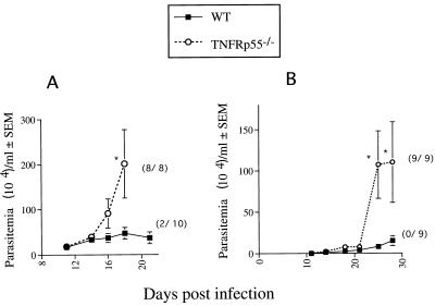
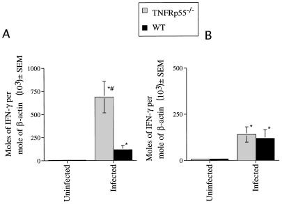
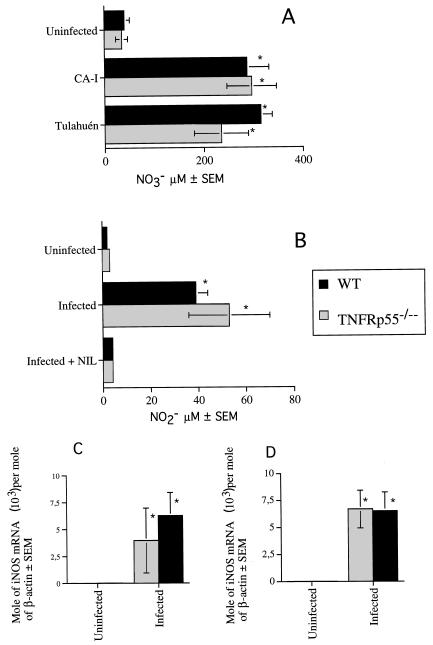
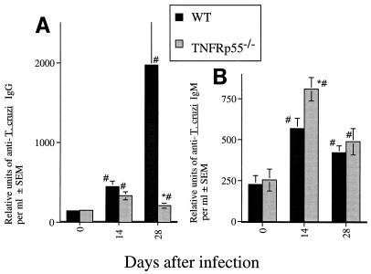
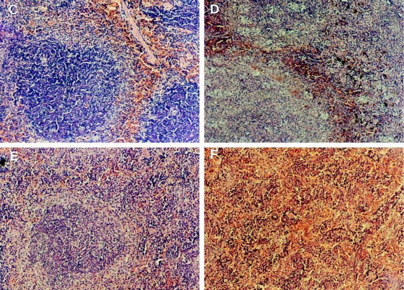
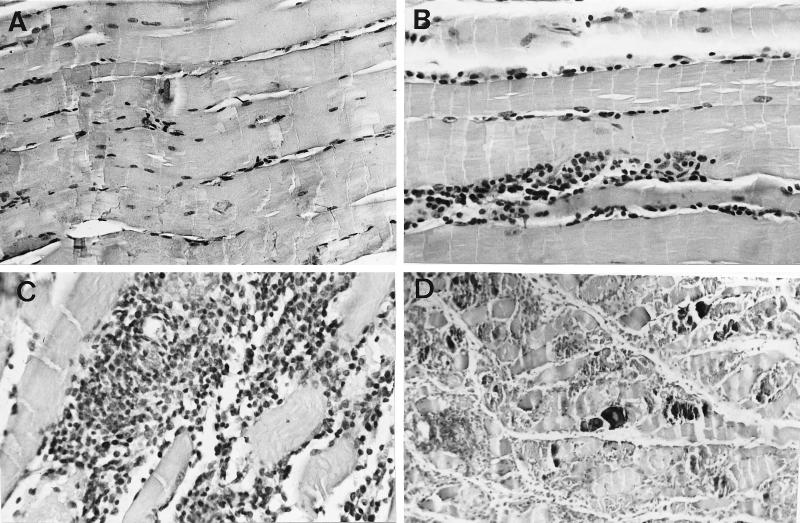
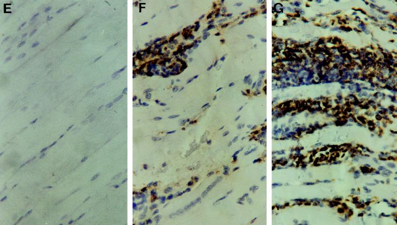
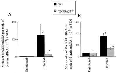
Tumor necrosis factor receptor p55 (TNFRp55) mediates host resistance to several pathogens by allowing microbicidal activities of phagocytes. In the studies reported here, TNFRp55-/- mice infected with the intracellular parasite Trypanosoma cruzi showed clearly higher parasitemia and cumulative mortality than wild-type (WT) controls did. However, gamma interferon (IFN-gamma)-activated macrophages from TNFRp55-/- mice produced control levels of nitric oxide and killed the parasite efficiently in vitro. Trypanocidal mechanisms of nonphagocytic cells (myocardial fibroblasts) from both TNFRp55-/- and WT mice were also activated by IFN-gamma in a dose-dependent way. However, IFN-gamma-activated TNFRp55-/- nonphagocytes showed less effective killing of T. cruzi than WT control nonphagocytes, even when interleukin 1beta (IL-1beta) was added as a costimulator. In vivo, T. cruzi-infected TNFRp55-/- mice and WT mice released similar levels of NO and showed similar levels of IFN-gamma mRNA and inducible nitric oxide synthase mRNA in their tissues. Instead, increased susceptibility to T. cruzi of TNFRp55-/- mice was associated with reduced levels of parasite-specific immunoglobulin G (IgG) (but not IgM) antibodies during infection, which is probably linked to abnormal B-cell differentiation in secondary lymphoid tissues of the mutant mice. Surprisingly, T. cruzi-infected TNFRp55-/- mice showed increased inflammatory and necrotic lesions in several tissues, especially in skeletal muscles, indicating that TNFRp55 plays an important role in controlling the inflammatory process. Accordingly, levels of Mn2+ superoxide dismutase mRNA, a TNF-induced enzyme which protects the cell from the toxic effects of superoxide, were lower in mutant than in WT infected mice.


Figures

FIG. 1
FIG. 1
Parasitemia and mortality of TNFRp55−/− and WT mice infected i.p. with 50 Tulahuén (A) or 104 CA-I (B) organisms. The mean number of organisms per milliliter ± standard error of the mean for one of two representative experiments with each parasite strain is depicted. The cumulative mortality at 20 (A) or 35 (B) days after infection is indicated in parentheses. Differences in cumulative mortality between WT and TNFRp55−/− mice were significant (P < 0.02, Fisher’s exact chi-square test). ∗, significantly different from the value for WT infected controls (P < 0.05, Mann-Whitney-U Wilcoxon test).
FIG. 2
FIG. 2
IFN-γ mRNA accumulation was determined in skeletal muscle (A) and spleens (B) from TNFRp55−/− and WT mice at 0 or 30 days after infection with T. cruzi (CA-I). Total RNA was obtained from spleens or skeletal muscle of individual mice, and IFN-γ and β-actin mRNA were measured in a competitive PCR assay. The mean moles of IFN-γ mRNA per mole of β-actin mRNA (used as a housekeeping gene) from four cDNA samples per group, of one of two independent experiments, are depicted. ∗, significantly different from the value for noninfected WT or mutant controls (P < 0.001, Mann-Whitney-U Wilcoxon test). #, significantly different from the value for WT infected mice (P < 0.05, Mann-Whitney-U Wilcoxon test).
FIG. 3
FIG. 3
Nitrate concentration in serum (A), nitrite content in cell supernatants (B), and iNOS mRNA accumulation in the spleen (C) and skeletal muscle (D) were determined in TNFRp55−/− and WT mice after infection with T. cruzi. Samples were obtained 20 and 30 days after infection with strains Tulahuén and CA-I, respectively. (A) The nitrate level was determined in serum from five individual mice for each group. The mean NO3 concentrations are depicted. ∗, significantly different from values for uninfected controls (P < 0.05, Mann-Whitney-U Wilcoxon test). (B) To measure NO release, peritoneal cells from individual mice (six per group) were cultured for 24 h in the presence or absence of 0.5 mM l-NIL. NO production was measured 24 h later by the Griess assay, and mean NO2 concentrations are depicted. The data of one of two independent experiments are shown. ∗, significantly different from value for uninfected mice (P < 0.05, Mann-Whitney-U Wilcoxon test). Differences between values for WT and TNFRp55−/− infected mice are not significant. (C and D) Total RNA was obtained from spleens (C) and skeletal muscle (D) of individual mice. iNOS and β-actin mRNA were measured in a competitive PCR assay. The mean moles of iNOS mRNA per mole of β-actin mRNA from four cDNA samples per group, of one of two independent experiments, are depicted. ∗, significantly different from value for uninfected controls (P < 0.05, Mann-Whitney-U Wilcoxon test).
FIG. 4
FIG. 4
(A and B) Titers of anti-T. cruzi IgG (A) and IgM (B) in sera from individual mice (six mice per group) at 17 and 28 days after infection with strain CA-I. The mean numbers of arbitrary units of specific IgG and IgM per milliliter obtained from one of two independent experiments are depicted. ∗, significantly different from values for WT infected mice (P < 0.05, F-test, analysis of variance). #, significantly different from values for uninfected WT or TNFRp55−/− controls (P < 0.05, F-test, analysis of variance). (C to F) Hematoxylin-eosin staining of spleen tissue sections from WT (C and D) and TNFRp55−/− mice (E and F) at 0 (C and E) or 15 (D and F) days after infection with T. cruzi. It is important to note the lack of primary follicles (E) and germinal center formation (F) in tissues from uninfected or infected TNFRp55−/− mice.
FIG. 4
FIG. 4
(A and B) Titers of anti-T. cruzi IgG (A) and IgM (B) in sera from individual mice (six mice per group) at 17 and 28 days after infection with strain CA-I. The mean numbers of arbitrary units of specific IgG and IgM per milliliter obtained from one of two independent experiments are depicted. ∗, significantly different from values for WT infected mice (P < 0.05, F-test, analysis of variance). #, significantly different from values for uninfected WT or TNFRp55−/− controls (P < 0.05, F-test, analysis of variance). (C to F) Hematoxylin-eosin staining of spleen tissue sections from WT (C and D) and TNFRp55−/− mice (E and F) at 0 (C and E) or 15 (D and F) days after infection with T. cruzi. It is important to note the lack of primary follicles (E) and germinal center formation (F) in tissues from uninfected or infected TNFRp55−/− mice.
FIG. 5
FIG. 5
Hematoxylin-eosin stainings (A to D) and immunohistochemical study of CD45 expression (E to G) of skeletal muscles from TNFRp55−/− (A, C, D, and G) and WT (B, E, and F) mice at 0 (A and E) or 30 (B, C, D, F, and G) days after infection with T. cruzi (strain CA-I). Negative controls for immunohistochemical stainings done by incubation with normal rat serum on tissues from T. cruzi-infected mice showed no peroxidase staining (data not shown). It is important to note the dramatically increased density of inflammatory infiltrates (C and G) and the presence of necrosis and calcification (D) in tissues obtained from infected TNFRp55−/− mice.
FIG. 5
FIG. 5
Hematoxylin-eosin stainings (A to D) and immunohistochemical study of CD45 expression (E to G) of skeletal muscles from TNFRp55−/− (A, C, D, and G) and WT (B, E, and F) mice at 0 (A and E) or 30 (B, C, D, F, and G) days after infection with T. cruzi (strain CA-I). Negative controls for immunohistochemical stainings done by incubation with normal rat serum on tissues from T. cruzi-infected mice showed no peroxidase staining (data not shown). It is important to note the dramatically increased density of inflammatory infiltrates (C and G) and the presence of necrosis and calcification (D) in tissues obtained from infected TNFRp55−/− mice.
FIG. 6
FIG. 6
MnSOD mRNA accumulation was determined in skeletal muscle (A) and spleens (B) from individual mice at 0 and 30 days after infection with T. cruzi (CA-I). MnSOD and β-actin mRNA were measured in a competitive PCR assay. The mean moles of MnSOD mRNA per mole of β-actin mRNA from four cDNA samples per group, of one of two independent experiments, are depicted. ∗, significantly different from value for infected WT mice (P < 0.05, Mann-Whitney-U Wilcoxon test). #, significantly different from value for uninfected controls (P < 0.05, Mann-Whitney-U Wilcoxon test).

References

    1. Bancroft G J, Sheehan K C F, Shreiber R D, Unanue E R. Tumour necrosis factor is involved in the T-cell independent pathway of macrophage activation in scid mice. J Immunol. 1989;143:127–135. - PubMed
    1. Bazzoni F, Beutler B. The tumor necrosis factor ligand and receptor families. N Engl J Med. 1996;334:1717–1725. - PubMed
    1. Black C M, Israelski D M, Suzuki Y, Remington J S. Effect of recombinant tumour necrosis factor on acute infection in mice with Toxoplasma gondii or Trypanosoma cruzi. Immunology. 1989;68:570–574. - PMC - PubMed
    1. Cardoni R L, Rottenberg M E, Segura E L. Increased production of reactive oxygen species by spleen cells from mice acutely infected with Trypanosoma cruzi. Cell Immunol. 1990;128:11–21. - PubMed
    1. Chandrasekar B, Melby P, Troyer D, Freeman G. Induction of proinflammatory cytokine expression in experimental acute chagasic cardiomyopathy. Biochem Biophys Res Commun. 1996;223:365–371. - PubMed

Publication types

MeSH terms