. 1998 Jun 15;509 ( Pt 3)(Pt 3):817-31.
doi: 10.1111/j.1469-7793.1998.817bm.x.

Potentiation of GABAergic synaptic transmission by AMPA receptors in mouse cerebellar stellate cells: changes during development

Affiliations

Potentiation of GABAergic synaptic transmission by AMPA receptors in mouse cerebellar stellate cells: changes during development

I Bureau et al. J Physiol. .

Abstract

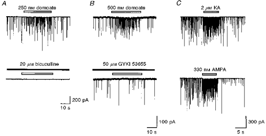
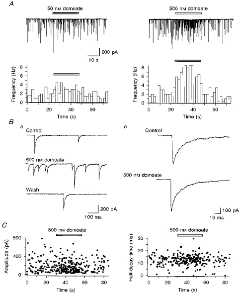
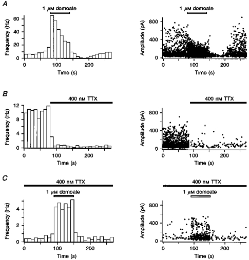
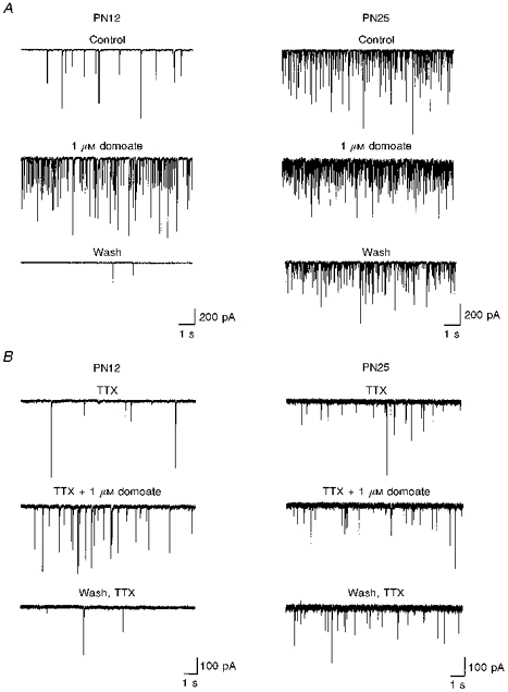
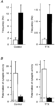
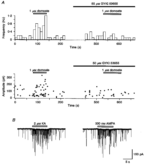
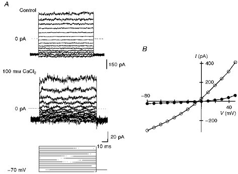
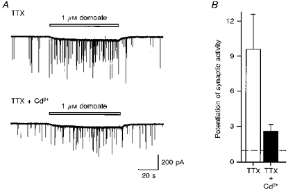
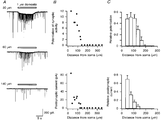
1. The effects of low concentrations of domoate, an agonist at both alpha-amino-3-hydroxy-5-methylisoxazole-4-propionate and kainate receptors (AMPARs and KARs, respectively), were investigated in stellate cells in slices of mouse cerebellum at two developmental stages (postnatal day (PN) 11-13 and PN21-25). 2. Low concentrations of domoate enhanced the frequency of miniature IPSCs (mIPSCs) recorded in the presence of tetrodotoxin (TTX) at PN11-13 but not at PN21-25. 3. The effects of low concentrations of domoate on synaptic activity were probably mediated by the activation of AMPARs and not KARs, since they were blocked by GYKI 53655 (LY300168), a selective AMPAR antagonist. 4. Domoate increased mIPSC frequency in part by activation of presynaptic voltage-dependent Ca2+ channels since potentiation was reduced by 60 % in the presence of Cd2+. AMPARs in stellate cells were found to be permeable to Ca2+. The residual potentiation in the presence of Cd2+ could thus be due to a direct entry of Ca2+ through AMPAR channels. 5. In the presence of TTX, potentiation of synaptic activity by focal application of domoate was not restricted to the region of the cell body, but was observed within distances of 120 micro(m). These experiments also revealed a strong spatial correlation between the location of the presynaptic effects of domoate and the activation of postsynaptic AMPARs. 6. Our data show a developmentally regulated presynaptic potentiation of synaptic transmission between cerebellar interneurones mediated by AMPARs. We discuss the possibility that the developmental switch could be due to a shift in the localization of AMPARs from the axonal to the somato-dendritic compartment.


Figures

Figure 2
Figure 2. Pharmacology of the effects of domoate on synaptic activity
A, samples of a continuous recording of spontaneous PSCs in a stellate cell. Bicuculline (20 μM) dramatically decreased spontaneous synaptic activity. In the presence of bicuculline (indicated by the filled horizontal bar), no potentiation of synaptic activity was induced by the application of 250 nM domoate (indicated by the open horizontal bars). B, addition of GYKI 53655 (50 μM; indicated by the filled horizontal bar) blocked the increase in spontaneous PSC frequency induced by 500 nM domoate. C, addition of KA (2 μM) and AMPA (330 nM) (indicated by the horizontal bars) induced a reversible potentiation of synaptic activity. Recordings in A-C are from 3 differents cells from PN14, PN14 and PN13 mice, respectively.
Figure 1
Figure 1. Low concentrations of domoate enhance synaptic activity in cerebellar stellate cells
A, upper panels: samples of a continuous recording of spontaneous PSCs in a stellate cell. Lower panels: plots for the same cell of the mean frequency of PSCs detected during 2 s sample intervals against time. Domoate (50 and 500 nM), applied during the time indicated by the horizontal bars, led to a reversible increase in synaptic activity in a dose-dependent manner. Ba and Bb, traces displaying, on an expanded time scale, selected portions of the data sample in the right-hand recording in A. Only PSCs with a long time course of decay, presumably GABAergic IPSCs, were observed. C, amplitude and half-decay time of synaptic events detected in continuous recordings in another cell. Domoate changed the frequency of the synaptic currents without obvious change in the amplitude or the time course of decay. Horizontal bars indicate the time during which 500 nM domoate was applied. Membrane holding potential was -70 mV. All experiments were performed using cells from PN17 mice.
Figure 3
Figure 3. Domoate increases the frequency of miniature IPSCs (mIPSCs) in stellate cells
Left panels: plots of the mean frequency of synaptic events detected during 10 s intervals against time. Right panels: plots of the amplitude of single IPSCs recorded continuously. All data are from the same neurone from a PN12 mouse. A, addition of domoate (1 μM) induced a large increase in spontaneous IPSC frequency which was followed, in this neurone, by a reversible decrease in synaptic activity as well as in IPSC amplitude that outlasted the application of domoate. B, in the same cell, addition of 400 nM TTX dramatically decreased spontaneous synaptic activity. C, in the presence of TTX, domoate induced an increase in the frequency of mIPSCs. The slice was bathed in physiological saline supplemented with 400 nM TTX.
Figure 4
Figure 4. Changes in spontaneous and mIPSC frequency and in the effect of domoate with age
Left panels: samples of a continuous recording of IPSCs in a stellate cell in the cerebellum from a PN12 mouse. Right panels: samples of a continuous recording of IPSCs in a stellate cell in the cerebellum from a PN25 mouse. In A, the slices were bathed in physiological saline. In B, the slices were bathed in physiological saline supplemented with 500 nM TTX.
Figure 5
Figure 5. Summary plots of the developmental changes in GABAergic synaptic activity and in the effects of domoate
A, pooled data (means ± s.e.m.) of the mean frequency of IPSCs under control conditions (left) and in the presence of 400-500 nM TTX (right). B, pooled data of the potentiation of synaptic activity induced by 1 μM domoate in stellate cells under control conditions (left) and in the presence of 400-500 nM TTX (right). Mean values ± s.e.m. of the ratio of mean frequency in the test condition to that in the control are shown. In A and B: □, PN11-13 mice (n = 6 for control conditions, n = 13 for experiments with TTX); ▪, PN21-25 mice (n = 7).
Figure 6
Figure 6. TTX-resistant effect of domoate on synaptic activity is blocked by GYKI 53655 and is mimicked by effects of KA and AMPA
A, upper panel: plots of the mean frequency of synaptic events detected during 10 s intervals against time. The potentiation of frequency of mIPSCs induced by 1 μM domoate was inhibited by 50 μM GYKI 53655. Lower panel: for the same cell, plots of the amplitude of each synaptic event recorded continuously. Open and filled horizontal bars indicate the time during which 1 μM domoate and 50 μM GYKI 53655 were applied, respectively. B, samples of a continuous recording of mIPSCs in a stellate cell. KA (2 μM) and AMPA (330 nM), applied during the time indicated by the horizontal bars, led to a reversible increase in synaptic activity in the same cell. The slice was bathed in physiological saline supplemented with 500 nM TTX. Cells from PN13 and PN12 mice were used in A and B, respectively.
Figure 7
Figure 7. Ca2+ permeability of AMPAR channels in stellate cells
A, domoate-activated currents recorded in a nucleated patch from a stellate cell, in control (high Na+; top traces) and in 100 mM CaCl2 (middle traces). For each voltage step, current traces were generated by subtracting the traces obtained under control conditions from those obtained during application of 50 μM domoate. The voltage protocol (10 mV steps) is shown at the bottom. B, I-V relations of currents activated by 50 μM domoate. These data are the graphic representation of currents shown in A. ○, control conditions (high Na+); •, in 100 mM CaCl2 (Na+-free extracellular solution). These experiments were performed using cells from a PN13 mouse.
Figure 8
Figure 8. CdCl2 partially blocks the potentiation of mIPSC frequency induced by domoate in stellate cells
A, samples of a continuous recording of mIPSCs in a stellate cell. CdCl2 (100 μM) decreased the effect of 1 μM domoate, applied during the time indicated by the horizontal bars, on mIPSC frequency without affecting the direct postsynaptic current. B, pooled data of the potentiation of synaptic activity induced by 1 μM domoate in stellate cells. □, data from experiments carried out in the presence of 500 nM TTX; ▪, data from experiments carried out in the presence of 500 nM TTX and 100 μM CdCl2 (n = 10). Mean values ± s.e.m. of the ratio of mean frequency in the test condition to that in the control are shown. All experiments were performed using cells from PN11-13 mice.
Figure 9
Figure 9. Focal application of domoate increases the frequency of mIPSCs in stellate cells
A, samples of a continuous recording of mIPSCs in a stellate cell in the cerebellum from a PN13 mouse. In the presence of TTX (500 nM), focal application of 1 μM domoate, indicated by the horizontal bars above the traces, increased mIPSC frequency when applied at a distance of 30 μm from the soma of the recorded stellate cell. This effect was decreased at 60 μm and was not observed at 140 μm. B shows, in the same cell, plots of the effects of focal application of domoate on mIPSC frequency, as measured by the ratio of mIPSC frequency in the test condition to that in the control (top), and postsynaptic current (bottom), as a function of the distance from the recorded stellate cell. C, pooled data for the effects of focal application of domoate (1 μM) on mIPSC frequency (top) and postsynaptic current (bottom). On the ordinate, values are given relative to the maximal potentiation and maximal postsynaptic current measured for each cell. All experiments were performed using cells from PN11-13 mice (n = 15).

Similar articles

Cited by

References

    1. Altman J, Bayer SA. Development of the Cerebellar System in Relation to its Evolution, Structure and Functions. Boca Raton, FL, USA: CRC Press; 1997.
    1. Ankri N, Legendre P, Faber DS, Korn H. Automatic detection of spontaneous synaptic responses in central neurones. Journal of Neuroscience Methods. 1994;52:87–100. 10.1016/0165-0270(94)90060-4. - DOI - PubMed
    1. Baude A, Molnar E, Latawiec D, McIlhinney RA, Somogyi P. Synaptic and nonsynaptic localization of the GluR1 subunit of the AMPA-type excitatory amino acid receptor in the rat cerebellum. Journal of Neuroscience. 1994;14:2830–2843. - PMC - PubMed
    1. Bettler B, Egebjerg J, Sharma G, Pecht G, Hermans-Borgmeyer I, Moll C, Stevens CF, Heinemann S. Cloning of a putative glutamate receptor: a low affinity kainate receptor subunit. Neuron. 1992;8:257–265. - PubMed
    1. Bettler B, Mulle C. AMPA and kainate receptors. Neuropharmacology. 1995;34:123–139. - PubMed

MeSH terms

LinkOut - more resources